 Movie
Movie Controller

[English] 日本語
 Yorodumi
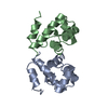
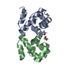
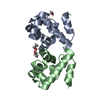
- PDB-1ci4: THE CRYSTAL STRUCTURE OF HUMAN BARRIER-TO-AUTOINTEGRATION FACTOR (BAF) -
+ Open data

- Basic information
| Entry | Database: PDB / ID: 1ci4 | |||||||||
|---|---|---|---|---|---|---|---|---|---|---|
| Title | THE CRYSTAL STRUCTURE OF HUMAN BARRIER-TO-AUTOINTEGRATION FACTOR (BAF) | |||||||||
 Components Components | PROTEIN (BARRIER-TO-AUTOINTEGRATION FACTOR (BAF)) | |||||||||
 Keywords Keywords | DNA BINDING PROTEIN / RETROVIRAL INTEGRATION / PREINTEGRATION COMPLEX | |||||||||
| Function / homology |  Function and homology information Function and homology informationnegative regulation of protein ADP-ribosylation / Nuclear Envelope Breakdown / mitotic nuclear membrane reassembly / Initiation of Nuclear Envelope (NE) Reformation / Integration of viral DNA into host genomic DNA / Autointegration results in viral DNA circles / negative regulation of viral genome replication / 2-LTR circle formation / negative regulation of cGAS/STING signaling pathway / Vpr-mediated nuclear import of PICs ...negative regulation of protein ADP-ribosylation / Nuclear Envelope Breakdown / mitotic nuclear membrane reassembly / Initiation of Nuclear Envelope (NE) Reformation / Integration of viral DNA into host genomic DNA / Autointegration results in viral DNA circles / negative regulation of viral genome replication / 2-LTR circle formation / negative regulation of cGAS/STING signaling pathway / Vpr-mediated nuclear import of PICs / negative regulation of type I interferon production / Integration of provirus / APOBEC3G mediated resistance to HIV-1 infection / chromosome organization / condensed chromosome / negative regulation of innate immune response / response to virus / nuclear envelope / chromatin organization / double-stranded DNA binding / response to oxidative stress / chromatin / protein homodimerization activity / DNA binding / nucleoplasm / identical protein binding / nucleus / cytosol / cytoplasm Similarity search - Function | |||||||||
| Biological species |  Homo sapiens (human) Homo sapiens (human) | |||||||||
| Method |  X-RAY DIFFRACTION / SYNCHROTRON / MAD / Resolution: 1.9 Å X-RAY DIFFRACTION / SYNCHROTRON / MAD / Resolution: 1.9 Å | |||||||||
 Authors Authors | Umland, T.C. / Wei, S.-Q. / Craigie, R. / Davies, D.R. | |||||||||
 Citation Citation | Journal: Biochemistry / Year: 2000 Title: Structural basis of DNA bridging by barrier-to-autointegration factor. Authors: Umland, T.C. / Wei, S.Q. / Craigie, R. / Davies, D.R. #1: Journal: Proc.Natl.Acad.Sci.USA / Year: 1998 Title: A previously unidentified host protein protects retroviral DNA from autointegration. Authors: Lee, M.S. / Craigie, R. #2: Journal: Proc.Natl.Acad.Sci.USA / Year: 1994 Title: Protection of retroviral DNA from autointegration: involvement of a cellular factor. Authors: Lee, M.S. / Craigie, R. | |||||||||
| History |
|
- Structure visualization
| Structure viewer | Molecule: MolmilJmol/JSmol |
|---|
- Downloads & links
-Download
| PDBx/mmCIF format | 1ci4.cif.gz | 52.3 KB | Display | PDBx/mmCIF format |
|---|---|---|---|---|
| PDB format | pdb1ci4.ent.gz | 37.3 KB | Display | PDB format |
| PDBx/mmJSON format | 1ci4.json.gz | Tree view | PDBx/mmJSON format | |
| Others |  Other downloads Other downloads |
-Validation report
| Arichive directory | https://data.pdbj.org/pub/pdb/validation_reports/ci/1ci4ftp://data.pdbj.org/pub/pdb/validation_reports/ci/1ci4 | HTTPS FTP |
|---|
-Related structure data
| Similar structure data |
|---|


-Links
 PDBj
PDBj


- Assembly
| Deposited unit | 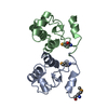
| ||||||||||||||||||||||||
|---|---|---|---|---|---|---|---|---|---|---|---|---|---|---|---|---|---|---|---|---|---|---|---|---|---|
| 1 |
| ||||||||||||||||||||||||
| Unit cell |
| ||||||||||||||||||||||||
| Components on special symmetry positions |
| ||||||||||||||||||||||||
| Noncrystallographic symmetry (NCS) | NCS domain:
NCS oper:
|
-Components
| #1: Protein | Mass: 10167.377 Da / Num. of mol.: 2 Source method: isolated from a genetically manipulated source Details: SELENOMETHIONINE (RESIDUE NAME MSE) HAS BEEN SUBSTITUTED FOR METHIONINE Source: (gene. exp.) Homo sapiens (human) / Plasmid: PET15B / Production host:  #2: Water | ChemComp-HOH / |  Mass: 18.015 Da / Num. of mol.: 228 / Source method: isolated from a natural source / Formula: H2O Mass: 18.015 Da / Num. of mol.: 228 / Source method: isolated from a natural source / Formula: H2OHas protein modification | Y | |
|---|
-Experimental details
-Experiment
| Experiment | Method: X-RAY DIFFRACTION / Number of used crystals: 1 |
|---|
- Sample preparation
| Crystal | Density Matthews: 2.3 Å3/Da / Density % sol: 44 % | ||||||||||||||||||||||||
|---|---|---|---|---|---|---|---|---|---|---|---|---|---|---|---|---|---|---|---|---|---|---|---|---|---|
| Crystal grow | pH: 6.5 Details: DIALYSIS OF PROTEIN AT 8.3 MG/ML IN 20MM TRIS HCL AT PH7.0, 10%(W/V) GLYCEROL, 150MM NACL, 10MM DTT, AND 0.1MM EDTA AGAINST 20MM IMIDAZOLE AT PH 6.5, 80MM NACL, AND 10MM DTT. DIALYSIS DONE ...Details: DIALYSIS OF PROTEIN AT 8.3 MG/ML IN 20MM TRIS HCL AT PH7.0, 10%(W/V) GLYCEROL, 150MM NACL, 10MM DTT, AND 0.1MM EDTA AGAINST 20MM IMIDAZOLE AT PH 6.5, 80MM NACL, AND 10MM DTT. DIALYSIS DONE AT ROOM TEMPERATURE.(ALL CONCENTRATIONS ARE IN MILLI-MOLAR) | ||||||||||||||||||||||||
| Crystal grow | *PLUS Method: microdialysis | ||||||||||||||||||||||||
| Components of the solutions | *PLUS
|
-Data collection
| Diffraction | Mean temperature: 95 K | ||||||||||||
|---|---|---|---|---|---|---|---|---|---|---|---|---|---|
| Diffraction source | Source: SYNCHROTRON / Site: NSLS  / Beamline: X9B / Wavelength: 0.9793,0.9789,0.9686 / Beamline: X9B / Wavelength: 0.9793,0.9789,0.9686 | ||||||||||||
| Detector | Type: MAR scanner 345 mm plate / Detector: IMAGE PLATE / Date: Jul 27, 1998 / Details: BENT MIRRORS | ||||||||||||
| Radiation | Monochromator: SI(111) / Protocol: MAD / Monochromatic (M) / Laue (L): M / Scattering type: x-ray | ||||||||||||
| Radiation wavelength |
| ||||||||||||
| Reflection | Resolution: 1.9→40 Å / Num. obs: 16011 / % possible obs: 99.5 % / Observed criterion σ(I): -3 / Redundancy: 13.7 % / Biso Wilson estimate: 17.3 Å2 / Rsym value: 6.1 / Net I/σ(I): 16 | ||||||||||||
| Reflection shell | Resolution: 1.9→1.94 Å / Rsym value: 18.4 / % possible all: 100 | ||||||||||||
| Reflection | *PLUS Num. measured all: 219206 / Rmerge(I) obs: 0.061 | ||||||||||||
| Reflection shell | *PLUS % possible obs: 100 % / Rmerge(I) obs: 0.184 |
- Processing
| Software |
| ||||||||||||||||||||||||||||||||||||||||||||||||||||||||||||||||||||||||||||||||
|---|---|---|---|---|---|---|---|---|---|---|---|---|---|---|---|---|---|---|---|---|---|---|---|---|---|---|---|---|---|---|---|---|---|---|---|---|---|---|---|---|---|---|---|---|---|---|---|---|---|---|---|---|---|---|---|---|---|---|---|---|---|---|---|---|---|---|---|---|---|---|---|---|---|---|---|---|---|---|---|---|---|
| Refinement | Method to determine structure: MAD / Resolution: 1.9→40 Å / Rfactor Rfree error: 0.007 / Data cutoff high rms absF: 281646.63 / Isotropic thermal model: RESTRAINED / Cross valid method: THROUGHOUT / σ(F): 0 / Stereochemistry target values: MLI Details: REFINEMENT TARGET: MAXIMUM LIKELIHOOD TARGET USING INTENSITIES
| ||||||||||||||||||||||||||||||||||||||||||||||||||||||||||||||||||||||||||||||||
| Solvent computation | Solvent model: FLAT MODEL / Bsol: 36.72 Å2 / ksol: 0.351 e/Å3 | ||||||||||||||||||||||||||||||||||||||||||||||||||||||||||||||||||||||||||||||||
| Displacement parameters | Biso mean: 24.1 Å2
| ||||||||||||||||||||||||||||||||||||||||||||||||||||||||||||||||||||||||||||||||
| Refine analyze |
| ||||||||||||||||||||||||||||||||||||||||||||||||||||||||||||||||||||||||||||||||
| Refinement step | Cycle: LAST / Resolution: 1.9→40 Å
| ||||||||||||||||||||||||||||||||||||||||||||||||||||||||||||||||||||||||||||||||
| Refine LS restraints |
| ||||||||||||||||||||||||||||||||||||||||||||||||||||||||||||||||||||||||||||||||
| Refine LS restraints NCS | Refine-ID: X-RAY DIFFRACTION
| ||||||||||||||||||||||||||||||||||||||||||||||||||||||||||||||||||||||||||||||||
| LS refinement shell | Resolution: 1.9→2.02 Å / Rfactor Rfree error: 0.02 / Total num. of bins used: 6
| ||||||||||||||||||||||||||||||||||||||||||||||||||||||||||||||||||||||||||||||||
| Xplor file |
| ||||||||||||||||||||||||||||||||||||||||||||||||||||||||||||||||||||||||||||||||
| Software | *PLUS Name: CNS / Version: 0.4 / Classification: refinement | ||||||||||||||||||||||||||||||||||||||||||||||||||||||||||||||||||||||||||||||||
| Refinement | *PLUS Num. reflection obs: 14112 / Rfactor Rfree: 0.265 / Rfactor Rwork: 0.214 | ||||||||||||||||||||||||||||||||||||||||||||||||||||||||||||||||||||||||||||||||
| Solvent computation | *PLUS | ||||||||||||||||||||||||||||||||||||||||||||||||||||||||||||||||||||||||||||||||
| Displacement parameters | *PLUS | ||||||||||||||||||||||||||||||||||||||||||||||||||||||||||||||||||||||||||||||||
| Refine LS restraints | *PLUS
|
