 Movie
Movie Controller

[English] 日本語
 Yorodumi
- PDB-1cf2: THREE-DIMENSIONAL STRUCTURE OF D-GLYCERALDEHYDE-3-PHOSPHATE DEHYD... -
+ Open data

- Basic information
| Entry | Database: PDB / ID: 1cf2 | ||||||
|---|---|---|---|---|---|---|---|
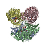
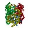
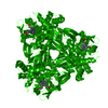
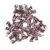
| Title | THREE-DIMENSIONAL STRUCTURE OF D-GLYCERALDEHYDE-3-PHOSPHATE DEHYDROGENASE FROM THE HYPERTHERMOPHILIC ARCHAEON METHANOTHERMUS FERVIDUS | ||||||
 Components Components | PROTEIN (GLYCERALDEHYDE-3-PHOSPHATE DEHYDROGENASE) | ||||||
 Keywords Keywords | OXIDOREDUCTASE / OXYDOREDUCTASE | ||||||
| Function / homology |  Function and homology information Function and homology informationglyceraldehyde-3-phosphate dehydrogenase (NAD(P)+) (phosphorylating) / 4-hydroxy-tetrahydrodipicolinate reductase activity / glyceraldehyde-3-phosphate dehydrogenase (NADP+) (phosphorylating) activity / glyceraldehyde-3-phosphate dehydrogenase (NAD+) (phosphorylating) activity / : / glycolytic process / NAD binding / NADP binding / cytoplasm Similarity search - Function | ||||||
| Biological species |   Methanothermus fervidus (archaea) Methanothermus fervidus (archaea) | ||||||
| Method |  X-RAY DIFFRACTION / SYNCHROTRON / MOLECULAR REPLACEMENT / Resolution: 2.1 Å X-RAY DIFFRACTION / SYNCHROTRON / MOLECULAR REPLACEMENT / Resolution: 2.1 Å | ||||||
 Authors Authors | Charron, C. / Talfournier, F. / Isuppov, M.N. / Branlant, G. / Littlechild, J.A. / Vitoux, B. / Aubry, A. | ||||||
 Citation Citation | Journal: Acta Crystallogr.,Sect.D / Year: 1999 Title: Crystallization and preliminary X-ray diffraction studies of D-glyceraldehyde-3-phosphate dehydrogenase from the hyperthermophilic archaeon Methanothermus fervidus. Authors: Charron, C. / Talfournier, F. / Isupov, M.N. / Branlant, G. / Littlechild, J.A. / Vitoux, B. / Aubry, A. | ||||||
| History |
|
- Structure visualization
| Structure viewer | Molecule: MolmilJmol/JSmol |
|---|
- Downloads & links
-Download
| PDBx/mmCIF format | 1cf2.cif.gz | 288.9 KB | Display | PDBx/mmCIF format |
|---|---|---|---|---|
| PDB format | pdb1cf2.ent.gz | 235.9 KB | Display | PDB format |
| PDBx/mmJSON format | 1cf2.json.gz | Tree view | PDBx/mmJSON format | |
| Others |  Other downloads Other downloads |
-Validation report
| Arichive directory | https://data.pdbj.org/pub/pdb/validation_reports/cf/1cf2ftp://data.pdbj.org/pub/pdb/validation_reports/cf/1cf2 | HTTPS FTP |
|---|
-Related structure data
| Similar structure data |
|---|


-Links
 PDBj
PDBj
- Assembly
| Deposited unit | 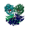
| ||||||||||||||||
|---|---|---|---|---|---|---|---|---|---|---|---|---|---|---|---|---|---|
| 1 |
| ||||||||||||||||
| Unit cell |
| ||||||||||||||||
| Noncrystallographic symmetry (NCS) | NCS oper:
|
-Components
| #1: Protein | Mass: 37454.793 Da / Num. of mol.: 4 Source method: isolated from a genetically manipulated source Source: (gene. exp.) Methanothermus fervidus (archaea) / Production host:  References: UniProt: P10618, glyceraldehyde-3-phosphate dehydrogenase (phosphorylating) #2: Chemical | ChemComp-SO4 /   Mass: 96.063 Da / Num. of mol.: 4 / Source method: obtained synthetically / Formula: SO4 Mass: 96.063 Da / Num. of mol.: 4 / Source method: obtained synthetically / Formula: SO4#3: Chemical | ChemComp-NAP /   Mass: 743.405 Da / Num. of mol.: 4 / Source method: obtained synthetically / Formula: C21H28N7O17P3 Mass: 743.405 Da / Num. of mol.: 4 / Source method: obtained synthetically / Formula: C21H28N7O17P3#4: Water | ChemComp-HOH / |  Mass: 18.015 Da / Num. of mol.: 960 / Source method: isolated from a natural source / Formula: H2O Mass: 18.015 Da / Num. of mol.: 960 / Source method: isolated from a natural source / Formula: H2O |
|---|
-Experimental details
-Experiment
| Experiment | Method: X-RAY DIFFRACTION / Number of used crystals: 1 |
|---|
- Sample preparation
| Crystal | Density Matthews: 2.62 Å3/Da / Density % sol: 53.01 % |
|---|---|
| Crystal grow | pH: 6.5 / Details: pH 6.50 |
| Crystal grow | *PLUS Method: vapor diffusion, hanging dropDetails: Jancarik, J., (1991) J. Appl. Crystallogr., 24, 409. |
| Components of the solutions | *PLUS Conc.: 10 mg/ml / Common name: protein |
-Data collection
| Diffraction | Mean temperature: 100 K |
|---|---|
| Diffraction source | Source: SYNCHROTRON / Site: ESRF  / Beamline: BM02 / Wavelength: 0.987 / Beamline: BM02 / Wavelength: 0.987 |
| Radiation | Protocol: SINGLE WAVELENGTH / Monochromatic (M) / Laue (L): M / Scattering type: x-ray |
| Radiation wavelength | Wavelength: 0.987 Å / Relative weight: 1 |
| Reflection | Resolution: 2.11→10 Å / Num. obs: 82965 / % possible obs: 91.7 % / Observed criterion σ(I): 2 / Redundancy: 5 % / Rsym value: 5.6 / Net I/σ(I): 9.1 |
| Reflection | *PLUS Highest resolution: 2.1 Å / Lowest resolution: 10 Å / % possible obs: 91.7 % / Observed criterion σ(I): 2 / Redundancy: 5 % / Num. measured all: 415252 / Rmerge(I) obs: 0.056 |
| Reflection shell | *PLUS Highest resolution: 2.11 Å / Lowest resolution: 2.17 Å / % possible obs: 69.3 % / Rmerge(I) obs: 0.151 / Mean I/σ(I) obs: 4 |
- Processing
| Software | Name: REFMAC / Classification: refinement | |||||||||||||||||||||||||||||||||||||||||||||||||||||||||||||||
|---|---|---|---|---|---|---|---|---|---|---|---|---|---|---|---|---|---|---|---|---|---|---|---|---|---|---|---|---|---|---|---|---|---|---|---|---|---|---|---|---|---|---|---|---|---|---|---|---|---|---|---|---|---|---|---|---|---|---|---|---|---|---|---|---|
| Refinement | Method to determine structure: MOLECULAR REPLACEMENT / Resolution: 2.1→10 Å / σ(F): 0
| |||||||||||||||||||||||||||||||||||||||||||||||||||||||||||||||
| Refinement step | Cycle: LAST / Resolution: 2.1→10 Å
| |||||||||||||||||||||||||||||||||||||||||||||||||||||||||||||||
| Refine LS restraints |
| |||||||||||||||||||||||||||||||||||||||||||||||||||||||||||||||
| Refinement | *PLUS Highest resolution: 2.1 Å / Lowest resolution: 10 Å / Rfactor obs: 0.194 | |||||||||||||||||||||||||||||||||||||||||||||||||||||||||||||||
| Solvent computation | *PLUS | |||||||||||||||||||||||||||||||||||||||||||||||||||||||||||||||
| Displacement parameters | *PLUS |
