 Movie
Movie Controller


+ Open data

- Basic information
| Entry | Database: PDB / ID: 1b7g | ||||||
|---|---|---|---|---|---|---|---|
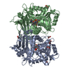
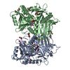
| Title | GLYCERALDEHYDE 3-PHOSPHATE DEHYDROGENASE | ||||||
 Components Components | PROTEIN (GLYCERALDEHYDE 3-PHOSPHATE DEHYDROGENASE) | ||||||
 Keywords Keywords | OXIDOREDUCTASE / ARCHAEA / HYPERTHERMOPHILE / GAPDH / HYPERTHERMOPHILIC DEHYDROGENASE | ||||||
| Function / homology |  Function and homology information Function and homology informationglyceraldehyde-3-phosphate dehydrogenase (NAD(P)+) (phosphorylating) / 4-hydroxy-tetrahydrodipicolinate reductase activity / glyceraldehyde-3-phosphate dehydrogenase (NADP+) (phosphorylating) activity / glyceraldehyde-3-phosphate dehydrogenase (NAD+) (phosphorylating) activity / : / glycolytic process / NAD binding / NADP binding / cytoplasm Similarity search - Function | ||||||
| Biological species |   Sulfolobus solfataricus (archaea) Sulfolobus solfataricus (archaea) | ||||||
| Method |  X-RAY DIFFRACTION / SYNCHROTRON / MIR / Resolution: 2.05 Å X-RAY DIFFRACTION / SYNCHROTRON / MIR / Resolution: 2.05 Å | ||||||
 Authors Authors | Isupov, M.N. / Littlechild, J.A. | ||||||
 Citation Citation | Journal: J.Mol.Biol. / Year: 1999 Title: Crystal structure of the glyceraldehyde-3-phosphate dehydrogenase from the hyperthermophilic archaeon Sulfolobus solfataricus. Authors: Isupov, M.N. / Fleming, T.M. / Dalby, A.R. / Crowhurst, G.S. / Bourne, P.C. / Littlechild, J.A. #1: Journal: Acta Crystallogr.,Sect.D / Year: 1998Title: Characterization, Crystallization and Preliminary X-Ray Investigation of Glyceraldehyde-3-Phosphate Dehydrogenase from the Hyperthermophilic Archaeon Sulfolobus Solfataricus Authors: Fleming, T.M. / Jones, C.E. / Piper, P.W. / Cowan, D.A. / Isupov, M.N. / Littlechild, J.A. | ||||||
| History |
|
- Structure visualization
| Structure viewer | Molecule: MolmilJmol/JSmol |
|---|
- Downloads & links
-Download
| PDBx/mmCIF format | 1b7g.cif.gz | 154.3 KB | Display | PDBx/mmCIF format |
|---|---|---|---|---|
| PDB format | pdb1b7g.ent.gz | 122.6 KB | Display | PDB format |
| PDBx/mmJSON format | 1b7g.json.gz | Tree view | PDBx/mmJSON format | |
| Others |  Other downloads Other downloads |
-Validation report
| Arichive directory | https://data.pdbj.org/pub/pdb/validation_reports/b7/1b7gftp://data.pdbj.org/pub/pdb/validation_reports/b7/1b7g | HTTPS FTP |
|---|
-Related structure data
| Similar structure data |
|---|


-Links
 PDBj
PDBj
- Assembly
| Deposited unit | 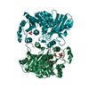
| ||||||||
|---|---|---|---|---|---|---|---|---|---|
| 1 |
| ||||||||
| 2 | 
| ||||||||
| Unit cell |
| ||||||||
| Noncrystallographic symmetry (NCS) | NCS oper: (Code: given Matrix: (-0.62399, 0.38067, 0.68244), Vector: |
-Components
| #1: Protein | Mass: 37631.617 Da / Num. of mol.: 2 / Mutation: YES Source method: isolated from a genetically manipulated source Source: (gene. exp.) Sulfolobus solfataricus (archaea) / Cellular location: CYTOPLASM / Production host:  References: UniProt: P39460, glyceraldehyde-3-phosphate dehydrogenase (phosphorylating) #2: Chemical | ChemComp-SO4 /   Mass: 96.063 Da / Num. of mol.: 12 / Source method: obtained synthetically / Formula: SO4 Mass: 96.063 Da / Num. of mol.: 12 / Source method: obtained synthetically / Formula: SO4#3: Water | ChemComp-HOH / |  Mass: 18.015 Da / Num. of mol.: 516 / Source method: isolated from a natural source / Formula: H2O Mass: 18.015 Da / Num. of mol.: 516 / Source method: isolated from a natural source / Formula: H2OHas protein modification | Y | Sequence details | VAL O 2, FOR EASE OF CLONING OF THE GENE IN E.COLI VAL Q 2, FOR EASE OF CLONING OF THE GENE IN E. ...VAL O 2, FOR EASE OF CLONING OF THE GENE IN E.COLI VAL Q 2, FOR EASE OF CLONING OF THE GENE IN E.COLI ILE O 48, CONFLICT BETWEEN THE TWO PUBLISHED SEQUENCES ILE Q 48, CONFLICT BETWEEN THE TWO PUBLISHED SEQUENCES ALA O 51, CONFLICT BETWEEN THE TWO PUBLISHED SEQUENCES ALA Q 51, CONFLICT BETWEEN THE TWO PUBLISHED SEQUENCES GLU O 72, CONFLICT BETWEEN THE TWO PUBLISHED SEQUENCES GLU Q 72, CONFLICT BETWEEN THE TWO PUBLISHED SEQUENCES ASP O 73, CONFLICT BETWEEN THE TWO PUBLISHED SEQUENCES ASP Q 73, CONFLICT BETWEEN THE TWO PUBLISHED SEQUENCES LYS O 159, CONFLICT BETWEEN THE TWO PUBLISHED SEQUENCES LYS Q 159, CONFLICT BETWEEN THE TWO PUBLISHED SEQUENCES | |
|---|
-Experimental details
-Experiment
| Experiment | Method: X-RAY DIFFRACTION / Number of used crystals: 1 |
|---|
- Sample preparation
| Crystal | Density Matthews: 3.1 Å3/Da / Density % sol: 60 % Description: THE STRUCTURE WAS SOLVED TO 3. ANGSTROM USING IN- HOUSE DATA | ||||||||||||||||||||||||||||||||||||||||
|---|---|---|---|---|---|---|---|---|---|---|---|---|---|---|---|---|---|---|---|---|---|---|---|---|---|---|---|---|---|---|---|---|---|---|---|---|---|---|---|---|---|
| Crystal grow | pH: 6.5 / Details: pH 6.50 | ||||||||||||||||||||||||||||||||||||||||
| Crystal | *PLUS Density % sol: 60 % | ||||||||||||||||||||||||||||||||||||||||
| Crystal grow | *PLUS pH: 6.5 / Method: vapor diffusion, hanging drop | ||||||||||||||||||||||||||||||||||||||||
| Components of the solutions | *PLUS
|
-Data collection
| Diffraction | Mean temperature: 100 K |
|---|---|
| Diffraction source | Source: SYNCHROTRON / Site: SRS  / Beamline: PX9.6 / Wavelength: 1 / Beamline: PX9.6 / Wavelength: 1 |
| Detector | Type: MARRESEARCH / Detector: IMAGE PLATE / Date: Aug 15, 1997 |
| Radiation | Protocol: SINGLE WAVELENGTH / Monochromatic (M) / Laue (L): M / Scattering type: x-ray |
| Radiation wavelength | Wavelength: 1 Å / Relative weight: 1 |
| Reflection | Resolution: 2.05→22 Å / Num. obs: 59606 / % possible obs: 99.6 % / Redundancy: 5.9 % / Biso Wilson estimate: 33 Å2 / Rmerge(I) obs: 0.056 / Net I/σ(I): 27.3 |
| Reflection shell | Resolution: 2.05→2.09 Å / Redundancy: 5.6 % / Rmerge(I) obs: 0.472 / Mean I/σ(I) obs: 2.2 / % possible all: 98.5 |
| Reflection | *PLUS Highest resolution: 2.05 Å / Lowest resolution: 22 Å / Redundancy: 5.9 % / Biso Wilson estimate: 33 Å2 |
| Reflection shell | *PLUS % possible obs: 98.5 % / Redundancy: 2.5 % / Mean I/σ(I) obs: 2.2 |
- Processing
| Software |
| ||||||||||||||||||||||||||||||||||||||||||||||||||||||||||||||||||||||||||||||||||||
|---|---|---|---|---|---|---|---|---|---|---|---|---|---|---|---|---|---|---|---|---|---|---|---|---|---|---|---|---|---|---|---|---|---|---|---|---|---|---|---|---|---|---|---|---|---|---|---|---|---|---|---|---|---|---|---|---|---|---|---|---|---|---|---|---|---|---|---|---|---|---|---|---|---|---|---|---|---|---|---|---|---|---|---|---|---|
| Refinement | Method to determine structure: MIR / Resolution: 2.05→22 Å / SU B: 5.4 / SU ML: 0.14 / Cross valid method: THROUGHOUT / σ(F): 0 / ESU R: 0.19 / ESU R Free: 0.19
| ||||||||||||||||||||||||||||||||||||||||||||||||||||||||||||||||||||||||||||||||||||
| Displacement parameters | Biso mean: 38 Å2
| ||||||||||||||||||||||||||||||||||||||||||||||||||||||||||||||||||||||||||||||||||||
| Refinement step | Cycle: LAST / Resolution: 2.05→22 Å
| ||||||||||||||||||||||||||||||||||||||||||||||||||||||||||||||||||||||||||||||||||||
| Refine LS restraints |
| ||||||||||||||||||||||||||||||||||||||||||||||||||||||||||||||||||||||||||||||||||||
| Software | *PLUS Name: REFMAC / Classification: refinement | ||||||||||||||||||||||||||||||||||||||||||||||||||||||||||||||||||||||||||||||||||||
| Refinement | *PLUS Rfactor obs: 0.229 | ||||||||||||||||||||||||||||||||||||||||||||||||||||||||||||||||||||||||||||||||||||
| Solvent computation | *PLUS | ||||||||||||||||||||||||||||||||||||||||||||||||||||||||||||||||||||||||||||||||||||
| Displacement parameters | *PLUS |
