 Movie
Movie Controller

[English] 日本語
 Yorodumi
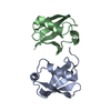
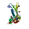
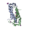
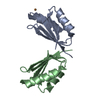
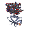
- PDB-1cc5: CRYSTAL STRUCTURE OF AZOTOBACTER CYTOCHROME C5 AT 2.5 ANGSTROMS R... -
+ Open data

- Basic information
| Entry | Database: PDB / ID: 1cc5 | ||||||
|---|---|---|---|---|---|---|---|
| Title | CRYSTAL STRUCTURE OF AZOTOBACTER CYTOCHROME C5 AT 2.5 ANGSTROMS RESOLUTION | ||||||
 Components Components | CYTOCHROME C5 | ||||||
 Keywords Keywords | ELECTRON TRANSPORT (HEME PROTEIN) | ||||||
| Function / homology |  Function and homology information Function and homology information | ||||||
| Biological species |  Azotobacter vinelandii (bacteria) Azotobacter vinelandii (bacteria) | ||||||
| Method |  X-RAY DIFFRACTION / Resolution: 2.5 Å X-RAY DIFFRACTION / Resolution: 2.5 Å | ||||||
 Authors Authors | Stout, C.D. / Carter, D.C. | ||||||
 Citation Citation | Journal: J.Mol.Biol. / Year: 1985 Title: Crystal structure of Azotobacter cytochrome c5 at 2.5 A resolution. Authors: Carter, D.C. / Melis, K.A. / O'Donnell, S.E. / Burgess, B.K. / Furey Jr., W.R. / Wang, B.C. / Stout, C.D. #1: Journal: J.Mol.Biol. / Year: 1978Title: Preliminary Crystallographic Data for Azotobacter Cytochrome C5 Authors: Stout, C.D. | ||||||
| History |
|
- Structure visualization
| Structure viewer | Molecule: MolmilJmol/JSmol |
|---|
- Downloads & links
-Download
| PDBx/mmCIF format | 1cc5.cif.gz | 27.9 KB | Display | PDBx/mmCIF format |
|---|---|---|---|---|
| PDB format | pdb1cc5.ent.gz | 17.3 KB | Display | PDB format |
| PDBx/mmJSON format | 1cc5.json.gz | Tree view | PDBx/mmJSON format | |
| Others |  Other downloads Other downloads |
-Validation report
| Arichive directory | https://data.pdbj.org/pub/pdb/validation_reports/cc/1cc5ftp://data.pdbj.org/pub/pdb/validation_reports/cc/1cc5 | HTTPS FTP |
|---|
-Related structure data
| Similar structure data |
|---|


-Links
 PDBj
PDBj


- Assembly
| Deposited unit | 
| ||||||||
|---|---|---|---|---|---|---|---|---|---|
| 1 | 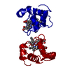
| ||||||||
| Unit cell |
| ||||||||
| Details | IN THIS SPACE GROUP, THE CYTOCHROME MOLECULE OCCUPIES A CRYSTALLOGRAPHIC TWO-FOLD SYMMETRY SITE. TO GENERATE THE DIMER THE FOLLOWING TRANSFORMATION SHOULD BE APPLIED TO THE COORDINATES GIVEN BELOW -1.0 0.0 0.0 -.36325 0.0 1.0 0.0 0.0 0.0 0.0 -1.0 .93169 IN THE FRACTIONAL COORDINATE FRAME THE SYMMETRY TRANSFORMATION IS GIVEN BY -1.0 0.0 0.0 0.0 0.0 1.0 0.0 0.0 0.0 0.0 -1.0 1.0 |
-Components
| #1: Protein | Mass: 8188.235 Da / Num. of mol.: 1 Source method: isolated from a genetically manipulated source Source: (gene. exp.) Azotobacter vinelandii (bacteria) / References: UniProt: P11732 |
|---|---|
| #2: Chemical | ChemComp-HEM /   Mass: 616.487 Da / Num. of mol.: 1 / Source method: obtained synthetically / Formula: C34H32FeN4O4 Mass: 616.487 Da / Num. of mol.: 1 / Source method: obtained synthetically / Formula: C34H32FeN4O4 |
| Has protein modification | Y |
-Experimental details
-Experiment
| Experiment | Method: X-RAY DIFFRACTION |
|---|
- Sample preparation
| Crystal | Density Matthews: 2.08 Å3/Da / Density % sol: 40.81 % | ||||||||||||||||||||
|---|---|---|---|---|---|---|---|---|---|---|---|---|---|---|---|---|---|---|---|---|---|
| Crystal grow | *PLUS pH: 6.9 / Method: vapor diffusion | ||||||||||||||||||||
| Components of the solutions | *PLUS
|
-Data collection
| Radiation | Scattering type: x-ray |
|---|---|
| Radiation wavelength | Relative weight: 1 |
| Reflection | *PLUS Highest resolution: 2.5 Å / Num. measured all: 11526 |
- Processing
| Refinement | Resolution: 2.5→10 Å / σ(F): 2 /
| ||||||||||||||||||||||||||||||||||||||||||||||||||||||||||||
|---|---|---|---|---|---|---|---|---|---|---|---|---|---|---|---|---|---|---|---|---|---|---|---|---|---|---|---|---|---|---|---|---|---|---|---|---|---|---|---|---|---|---|---|---|---|---|---|---|---|---|---|---|---|---|---|---|---|---|---|---|---|
| Refinement step | Cycle: LAST / Resolution: 2.5→10 Å
| ||||||||||||||||||||||||||||||||||||||||||||||||||||||||||||
| Refine LS restraints |
| ||||||||||||||||||||||||||||||||||||||||||||||||||||||||||||
| Refinement | *PLUS | ||||||||||||||||||||||||||||||||||||||||||||||||||||||||||||
| Solvent computation | *PLUS | ||||||||||||||||||||||||||||||||||||||||||||||||||||||||||||
| Displacement parameters | *PLUS Biso mean: 10.1 Å2 | ||||||||||||||||||||||||||||||||||||||||||||||||||||||||||||
| Refine LS restraints | *PLUS Type: o_planar_d / Dev ideal: 0.019 | ||||||||||||||||||||||||||||||||||||||||||||||||||||||||||||
| LS refinement shell | *PLUS Highest resolution: 2.5 Å / Lowest resolution: 2.7 Å / Num. reflection obs: 336 / Rfactor obs: 0.258 |
