 Movie
Movie Controller


+ Open data

- Basic information
| Entry | Database: PDB / ID: 1cbi | ||||||
|---|---|---|---|---|---|---|---|
| Title | APO-CELLULAR RETINOIC ACID BINDING PROTEIN I | ||||||
 Components Components | CELLULAR RETINOIC ACID BINDING PROTEIN I | ||||||
 Keywords Keywords | RETINOIC-ACID TRANSPORT | ||||||
| Function / homology |  Function and homology information Function and homology informationRA biosynthesis pathway / retinoid binding / retinoic acid binding / retinal binding / retinol binding / fatty acid transport / fatty acid binding / nucleus / cytoplasm / cytosol Similarity search - Function | ||||||
| Biological species |  | ||||||
| Method |  X-RAY DIFFRACTION / Resolution: 2.7 Å X-RAY DIFFRACTION / Resolution: 2.7 Å | ||||||
 Authors Authors | Thompson, J.R. / Bratt, J.M. / Banaszak, L.J. | ||||||
 Citation Citation | Journal: J.Mol.Biol. / Year: 1995 Title: Crystal structure of cellular retinoic acid binding protein I shows increased access to the binding cavity due to formation of an intermolecular beta-sheet. Authors: Thompson, J.R. / Bratt, J.M. / Banaszak, L.J. #1: Journal: Structure / Year: 1994Title: Crystal Structures of Cellular Retinoic Acid Binding Proteins I and II in Complex with All-Trans-Retinoic Acid and a Synthetic Retinoid Authors: Kleywegt, G.J. / Bergfors, T. / Senn, H. / Le Motte, P. / Gsell, B. / Shudo, K. / Jones, A.T. | ||||||
| History |
| ||||||
| Remark 700 | SHEET CRABPI IS ACTUALLY A TEN-STRANDED BETA-BARREL. THIS IS REPRESENTED IN SHEET RECORDS BELOW BY ...SHEET CRABPI IS ACTUALLY A TEN-STRANDED BETA-BARREL. THIS IS REPRESENTED IN SHEET RECORDS BELOW BY AN ELEVEN-STRANDED SHEET. |
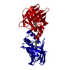
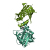
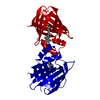
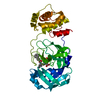
- Structure visualization
| Structure viewer | Molecule: MolmilJmol/JSmol |
|---|
- Downloads & links
-Download
| PDBx/mmCIF format | 1cbi.cif.gz | 75.4 KB | Display | PDBx/mmCIF format |
|---|---|---|---|---|
| PDB format | pdb1cbi.ent.gz | 57.7 KB | Display | PDB format |
| PDBx/mmJSON format | 1cbi.json.gz | Tree view | PDBx/mmJSON format | |
| Others |  Other downloads Other downloads |
-Validation report
| Arichive directory | https://data.pdbj.org/pub/pdb/validation_reports/cb/1cbiftp://data.pdbj.org/pub/pdb/validation_reports/cb/1cbi | HTTPS FTP |
|---|
-Related structure data
| Similar structure data |
|---|


-Links
 PDBj
PDBj

- Assembly
| Deposited unit | 
| ||||||||
|---|---|---|---|---|---|---|---|---|---|
| 1 |
| ||||||||
| Unit cell |
| ||||||||
| Noncrystallographic symmetry (NCS) | NCS oper: (Code: given Matrix: (0.8722, -0.4867, 0.0495), Vector: Details | MTRIX THE TRANSFORMATIONS PRESENTED ON MTRIX RECORDS BELOW DESCRIBE NON-CRYSTALLOGRAPHIC RELATIONSHIPS AMONG THE VARIOUS DOMAINS IN THIS ENTRY. APPLYING THE APPROPRIATE MTRIX TRANSFORMATION TO THE RESIDUES LISTED FIRST WILL YIELD APPROXIMATE COORDINATES FOR THE RESIDUES LISTED SECOND. APPLIED TO TRANSFORMED TO MTRIX RESIDUES RESIDUES RMSD | |
-Components
| #1: Protein | Mass: 15480.392 Da / Num. of mol.: 2 Source method: isolated from a genetically manipulated source Details: APO FORM / Source: (gene. exp.)  #2: Water | ChemComp-HOH / |  Mass: 18.015 Da / Num. of mol.: 71 / Source method: isolated from a natural source / Formula: H2O Mass: 18.015 Da / Num. of mol.: 71 / Source method: isolated from a natural source / Formula: H2O |
|---|
-Experimental details
-Experiment
| Experiment | Method: X-RAY DIFFRACTION |
|---|
- Sample preparation
| Crystal | Density Matthews: 4.06 Å3/Da / Density % sol: 69.69 % | ||||||||||||||||||||||||||||||
|---|---|---|---|---|---|---|---|---|---|---|---|---|---|---|---|---|---|---|---|---|---|---|---|---|---|---|---|---|---|---|---|
| Crystal grow | *PLUS Temperature: 18 ℃ / pH: 5 / Method: vapor diffusion, hanging drop | ||||||||||||||||||||||||||||||
| Components of the solutions | *PLUS
|
-Data collection
| Diffraction | Mean temperature: 294 K |
|---|---|
| Diffraction source | Wavelength: 1.5418 |
| Detector | Type: SIEMENS / Detector: AREA DETECTOR / Date: Nov 16, 1993 |
| Radiation | Monochromatic (M) / Laue (L): M / Scattering type: x-ray |
| Radiation wavelength | Wavelength: 1.5418 Å / Relative weight: 1 |
| Reflection | Num. obs: 12365 / % possible obs: 90.9 % / Observed criterion σ(I): 0 / Redundancy: 23 % / Rmerge(I) obs: 0.109 |
| Reflection | *PLUS Highest resolution: 2.7 Å / Lowest resolution: 13 Å / Rmerge(I) obs: 0.109 |
| Reflection shell | *PLUS Highest resolution: 2.7 Å / Lowest resolution: 2.9 Å / % possible obs: 51 % / Redundancy: 5.2 % / Rmerge(I) obs: 0.371 / Mean I/σ(I) obs: 1.69 |
- Processing
| Software |
| ||||||||||||||||||||||||||||||||||||||||||||||||||||||||||||
|---|---|---|---|---|---|---|---|---|---|---|---|---|---|---|---|---|---|---|---|---|---|---|---|---|---|---|---|---|---|---|---|---|---|---|---|---|---|---|---|---|---|---|---|---|---|---|---|---|---|---|---|---|---|---|---|---|---|---|---|---|---|
| Refinement | Resolution: 2.7→13 Å / σ(F): 2
| ||||||||||||||||||||||||||||||||||||||||||||||||||||||||||||
| Displacement parameters | Biso mean: 14 Å2 | ||||||||||||||||||||||||||||||||||||||||||||||||||||||||||||
| Refine analyze | Luzzati coordinate error obs: 0.3 Å | ||||||||||||||||||||||||||||||||||||||||||||||||||||||||||||
| Refinement step | Cycle: LAST / Resolution: 2.7→13 Å
| ||||||||||||||||||||||||||||||||||||||||||||||||||||||||||||
| Refine LS restraints |
| ||||||||||||||||||||||||||||||||||||||||||||||||||||||||||||
| Software | *PLUS Name: X-PLOR / Classification: refinement | ||||||||||||||||||||||||||||||||||||||||||||||||||||||||||||
| Refinement | *PLUS | ||||||||||||||||||||||||||||||||||||||||||||||||||||||||||||
| Solvent computation | *PLUS | ||||||||||||||||||||||||||||||||||||||||||||||||||||||||||||
| Displacement parameters | *PLUS | ||||||||||||||||||||||||||||||||||||||||||||||||||||||||||||
| Refine LS restraints | *PLUS
|
