 Movie
Movie Controller


+ Open data

- Basic information
| Entry | Database: PDB / ID: 1c76 | ||||||
|---|---|---|---|---|---|---|---|
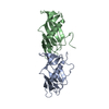
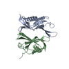
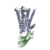
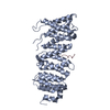
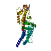
| Title | STAPHYLOKINASE (SAK) MONOMER | ||||||
 Components Components | STAPHYLOKINASE | ||||||
 Keywords Keywords | HYDROLASE / BETA-GRASP FAMILY | ||||||
| Function / homology |  Function and homology information Function and homology informationsymbiont-mediated activation of host plasminogen / symbiont-mediated suppression of host complement activation by activation of host proteases / extracellular region Similarity search - Function | ||||||
| Biological species |   Staphylococcus aureus (bacteria) Staphylococcus aureus (bacteria) | ||||||
| Method |  X-RAY DIFFRACTION / Resolution: 2.25 Å X-RAY DIFFRACTION / Resolution: 2.25 Å | ||||||
 Authors Authors | Rao, Z. / Jiang, F. / Liu, Y. / Zhang, X. / Chen, Y. / Bartlam, M. / Song, H. / Ding, Y. | ||||||
 Citation Citation | Journal: To be published Title: Crystal Structure of Staphylokinase Dimer Offers New Clue to Reduction of Immunogenicity Authors: Rao, Z. / Jiang, F. / Liu, Y. / Zhang, X. / Chen, Y. / Bartlam, M. / Song, H. / Ding, Y. | ||||||
| History |
|
- Structure visualization
| Structure viewer | Molecule: MolmilJmol/JSmol |
|---|
- Downloads & links
-Download
| PDBx/mmCIF format | 1c76.cif.gz | 35.7 KB | Display | PDBx/mmCIF format |
|---|---|---|---|---|
| PDB format | pdb1c76.ent.gz | 24.8 KB | Display | PDB format |
| PDBx/mmJSON format | 1c76.json.gz | Tree view | PDBx/mmJSON format | |
| Others |  Other downloads Other downloads |
-Validation report
| Arichive directory | https://data.pdbj.org/pub/pdb/validation_reports/c7/1c76ftp://data.pdbj.org/pub/pdb/validation_reports/c7/1c76 | HTTPS FTP |
|---|
-Related structure data
| Related structure data | |
|---|---|
| Similar structure data |


-Links
 PDBj
PDBj- Assembly
| Deposited unit | 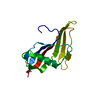
| ||||||||
|---|---|---|---|---|---|---|---|---|---|
| 1 | 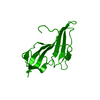
| ||||||||
| Unit cell |
| ||||||||
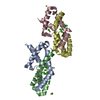
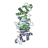
| Details | It is proposed in the primary citation that the biological unit is a "head-to-tail" dimer (pdb entry 1c77). Two other dimers have been considered, with alpha helices at the interface (1c78) and beta sheets at the interface (1c79) respectively. However, the "head-to-tail" dimer interface allows for the formation of strong hydrophobic interactions as well as hydrogen bonds and is more stable than the other two forms. The authors have experimental evidence from site direct mutagenesis which supports their proposition. |
-Components
| #1: Protein | Mass: 15487.585 Da / Num. of mol.: 1 Source method: isolated from a genetically manipulated source Source: (gene. exp.) Staphylococcus aureus (bacteria) / Production host: |
|---|---|
| #2: Water | ChemComp-HOH /  Mass: 18.015 Da / Num. of mol.: 16 / Source method: isolated from a natural source / Formula: H2O Mass: 18.015 Da / Num. of mol.: 16 / Source method: isolated from a natural source / Formula: H2O |
-Experimental details
-Experiment
| Experiment | Method: X-RAY DIFFRACTION / Number of used crystals: 1 |
|---|
- Sample preparation
| Crystal | Density Matthews: 2.18 Å3/Da / Density % sol: 43.6 % |
|---|---|
| Crystal grow | Temperature: 293 K / Method: vapor diffusion, hanging drop / pH: 8 Details: PEG 8000, pH 8.0, vapor diffusion/hanging drop, temperature 293K |
-Data collection
| Diffraction | Mean temperature: 298 K |
|---|---|
| Diffraction source | Source: ROTATING ANODE / Type: RIGAKU RU200 / Wavelength: 1.5418 |
| Detector | Type: MARRESEARCH / Detector: IMAGE PLATE / Date: Nov 28, 1999 |
| Radiation | Protocol: SINGLE WAVELENGTH / Monochromatic (M) / Laue (L): M / Scattering type: x-ray |
| Radiation wavelength | Wavelength: 1.5418 Å / Relative weight: 1 |
| Reflection | Resolution: 2.25→20 Å / Num. all: 6589 / Num. obs: 6589 / % possible obs: 97.8 % / Observed criterion σ(I): 0 / Redundancy: 4.4 % / Biso Wilson estimate: 20.5 Å2 / Rmerge(I) obs: 0.043 / Net I/σ(I): 12.8 |
| Reflection shell | Resolution: 2.25→2.35 Å / Redundancy: 4.8 % / Rmerge(I) obs: 0.182 / Num. unique all: 803 / % possible all: 97.6 |
- Processing
| Software |
| ||||||||||||||||||||
|---|---|---|---|---|---|---|---|---|---|---|---|---|---|---|---|---|---|---|---|---|---|
| Refinement | Resolution: 2.25→20 Å / σ(F): 2 / Stereochemistry target values: Engh & Huber
| ||||||||||||||||||||
| Refinement step | Cycle: LAST / Resolution: 2.25→20 Å
| ||||||||||||||||||||
| Refine LS restraints |
|
