 Movie
Movie Controller

[English] 日本語
 Yorodumi
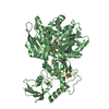
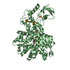
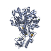
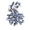
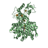
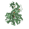
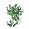
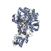
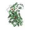
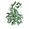
- PDB-1c4c: BINDING OF EXOGENOUSLY ADDED CARBON MONOXIDE AT THE ACTIVE SITE O... -
+ Open data

- Basic information
| Entry | Database: PDB / ID: 1c4c | ||||||
|---|---|---|---|---|---|---|---|
| Title | BINDING OF EXOGENOUSLY ADDED CARBON MONOXIDE AT THE ACTIVE SITE OF THE FE-ONLY HYDROGENASE (CPI) FROM CLOSTRIDIUM PASTEURIANUM | ||||||
 Components Components | PROTEIN (FE-ONLY HYDROGENASE) | ||||||
 Keywords Keywords | OXIDOREDUCTASE / METALLOPROTEINS / [FES] CLUSTERS / HYDROGEN OXIDATION / PROTON REDUCTION | ||||||
| Function / homology |  Function and homology information Function and homology informationferredoxin hydrogenase / ferredoxin hydrogenase activity / 4 iron, 4 sulfur cluster binding / iron ion binding Similarity search - Function | ||||||
| Biological species |  Clostridium pasteurianum (bacteria) Clostridium pasteurianum (bacteria) | ||||||
| Method |  X-RAY DIFFRACTION / MOLECULAR REPLACEMENT / Resolution: 2.4 Å X-RAY DIFFRACTION / MOLECULAR REPLACEMENT / Resolution: 2.4 Å | ||||||
 Authors Authors | Lemon, B.J. / Peters, J.W. | ||||||
 Citation Citation | Journal: Biochemistry / Year: 1999 Title: Binding of exogenously added carbon monoxide at the active site of the iron-only hydrogenase (CpI) from Clostridium pasteurianum. Authors: Lemon, B.J. / Peters, J.W. | ||||||
| History |
|
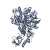
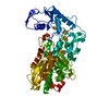
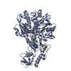
- Structure visualization
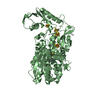
| Structure viewer | Molecule: MolmilJmol/JSmol |
|---|
- Downloads & links
-Download
| PDBx/mmCIF format | 1c4c.cif.gz | 133.2 KB | Display | PDBx/mmCIF format |
|---|---|---|---|---|
| PDB format | pdb1c4c.ent.gz | 102.3 KB | Display | PDB format |
| PDBx/mmJSON format | 1c4c.json.gz | Tree view | PDBx/mmJSON format | |
| Others |  Other downloads Other downloads |
-Validation report
| Arichive directory | https://data.pdbj.org/pub/pdb/validation_reports/c4/1c4cftp://data.pdbj.org/pub/pdb/validation_reports/c4/1c4c | HTTPS FTP |
|---|
-Related structure data
| Related structure data |  1c4aC  1fehS S: Starting model for refinement C: citing same article ( |
|---|---|
| Similar structure data |


-Links
 PDBj
PDBj


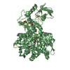
- Assembly
| Deposited unit | 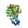
| ||||||||
|---|---|---|---|---|---|---|---|---|---|
| 1 |
| ||||||||
| Unit cell |
|
-Components
| #1: Protein | Mass: 63911.102 Da / Num. of mol.: 1 / Source method: isolated from a natural source / Source: (natural) Clostridium pasteurianum (bacteria) / Cellular location: CYTOPLASM / References: UniProt: P29166, 1.18.99.1 | ||||
|---|---|---|---|---|---|
| #2: Chemical | ChemComp-HC0 /   Mass: 366.936 Da / Num. of mol.: 1 / Source method: obtained synthetically / Formula: C6H7Fe2O7S2 Mass: 366.936 Da / Num. of mol.: 1 / Source method: obtained synthetically / Formula: C6H7Fe2O7S2 | ||||
| #3: Chemical | ChemComp-SF4 /   Mass: 351.640 Da / Num. of mol.: 4 / Source method: obtained synthetically / Formula: Fe4S4 Mass: 351.640 Da / Num. of mol.: 4 / Source method: obtained synthetically / Formula: Fe4S4#4: Chemical | ChemComp-FES / |   Mass: 175.820 Da / Num. of mol.: 1 / Source method: obtained synthetically / Formula: Fe2S2 Mass: 175.820 Da / Num. of mol.: 1 / Source method: obtained synthetically / Formula: Fe2S2#5: Water | ChemComp-HOH / |  Mass: 18.015 Da / Num. of mol.: 314 / Source method: isolated from a natural source / Formula: H2O Mass: 18.015 Da / Num. of mol.: 314 / Source method: isolated from a natural source / Formula: H2O |
-Experimental details
-Experiment
| Experiment | Method: X-RAY DIFFRACTION |
|---|
- Sample preparation
| Crystal | Density Matthews: 2.6 Å3/Da / Density % sol: 52.7 % | ||||||||||||||||||||
|---|---|---|---|---|---|---|---|---|---|---|---|---|---|---|---|---|---|---|---|---|---|
| Crystal grow | pH: 5 / Details: pH 5.0 | ||||||||||||||||||||
| Crystal grow | *PLUS pH: 4.6 / Method: batch method | ||||||||||||||||||||
| Components of the solutions | *PLUS
|
-Data collection
| Diffraction | Mean temperature: 100 K |
|---|---|
| Diffraction source | Source: ROTATING ANODE / Type: RIGAKU RUH3R / Wavelength: 1.5418 |
| Detector | Type: MACSCIENCE / Detector: IMAGE PLATE / Date: Apr 15, 1999 |
| Radiation | Protocol: SINGLE WAVELENGTH / Monochromatic (M) / Laue (L): M / Scattering type: x-ray |
| Radiation wavelength | Wavelength: 1.5418 Å / Relative weight: 1 |
| Reflection | Resolution: 2.4→20 Å / Num. obs: 24879 / % possible obs: 97 % / Observed criterion σ(I): 1 / Redundancy: 9 % / Rmerge(I) obs: 0.052 |
| Reflection shell | Resolution: 2.4→2.51 Å / Rmerge(I) obs: 0.13 / % possible all: 79.1 |
| Reflection | *PLUS Num. measured all: 234583 |
| Reflection shell | *PLUS % possible obs: 79.1 % |
- Processing
| Software |
| ||||||||||||||||||||||||||||||||||||||||||||||||||||||||||||
|---|---|---|---|---|---|---|---|---|---|---|---|---|---|---|---|---|---|---|---|---|---|---|---|---|---|---|---|---|---|---|---|---|---|---|---|---|---|---|---|---|---|---|---|---|---|---|---|---|---|---|---|---|---|---|---|---|---|---|---|---|---|
| Refinement | Method to determine structure: MOLECULAR REPLACEMENT Starting model: PDB ENTRY 1FEH Resolution: 2.4→20 Å / Cross valid method: THROUGHOUT / σ(F): 1
| ||||||||||||||||||||||||||||||||||||||||||||||||||||||||||||
| Refinement step | Cycle: LAST / Resolution: 2.4→20 Å
| ||||||||||||||||||||||||||||||||||||||||||||||||||||||||||||
| Refine LS restraints |
|
