- PDB-1c17: A1C12 SUBCOMPLEX OF F1FO ATP SYNTHASE -
+
Open data
ID or keywords:
Loading...
-
Basic information
Entry
Database: PDB / ID: 1c17
Title
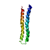
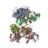
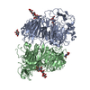
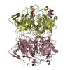
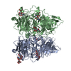
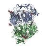
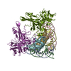
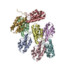
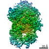
A1C12 SUBCOMPLEX OF F1FO ATP SYNTHASE
Components
ATP SYNTHASE SUBUNIT A
ATP SYNTHASE SUBUNIT C
Keywords
MEMBRANE PROTEIN / HELIX / COMPLEX
Function / homology
Function and homology information
proton motive force-driven plasma membrane ATP synthesis / proton motive force-driven ATP synthesis / proton-transporting two-sector ATPase complex, proton-transporting domain / proton-transporting ATPase activity, rotational mechanism / proton-transporting ATP synthase complex / proton-transporting ATP synthase activity, rotational mechanism / lipid binding / plasma membrane Similarity search - Function
ATP synthase, F0 complex, subunit A / F1F0 ATP synthase subunit C / F1FO ATP Synthase / ATP synthase, F0 complex, subunit A, bacterial/chloroplast / ATP synthase, F0 complex, subunit C, bacterial/chloroplast / ATP synthase, F0 complex, subunit A / ATP synthase, F0 complex, subunit A, active site / ATP synthase, F0 complex, subunit A superfamily / ATP synthase A chain / ATP synthase a subunit signature. ...ATP synthase, F0 complex, subunit A / F1F0 ATP synthase subunit C / F1FO ATP Synthase / ATP synthase, F0 complex, subunit A, bacterial/chloroplast / ATP synthase, F0 complex, subunit C, bacterial/chloroplast / ATP synthase, F0 complex, subunit A / ATP synthase, F0 complex, subunit A, active site / ATP synthase, F0 complex, subunit A superfamily / ATP synthase A chain / ATP synthase a subunit signature. / ATP synthase, F0 complex, subunit C / F1F0 ATP synthase subunit C superfamily / ATP synthase, F0 complex, subunit C, DCCD-binding site / ATP synthase c subunit signature. / V-ATPase proteolipid subunit C-like domain / F/V-ATP synthase subunit C superfamily / ATP synthase subunit C / Four Helix Bundle (Hemerythrin (Met), subunit A) / Up-down Bundle / Mainly Alpha Similarity search - Domain/homology
A: ATP SYNTHASE SUBUNIT C B: ATP SYNTHASE SUBUNIT C C: ATP SYNTHASE SUBUNIT C D: ATP SYNTHASE SUBUNIT C E: ATP SYNTHASE SUBUNIT C F: ATP SYNTHASE SUBUNIT C G: ATP SYNTHASE SUBUNIT C H: ATP SYNTHASE SUBUNIT C I: ATP SYNTHASE SUBUNIT C J: ATP SYNTHASE SUBUNIT C K: ATP SYNTHASE SUBUNIT C L: ATP SYNTHASE SUBUNIT C M: ATP SYNTHASE SUBUNIT A
Mass: 8259.064 Da / Num. of mol.: 12 Source method: isolated from a genetically manipulated source Source: (gene. exp.) Escherichia coli (E. coli) / Production host: Escherichia coli (E. coli) / References: UniProt: P68699
#2: Protein
ATPSYNTHASESUBUNITA
Mass: 19869.004 Da / Num. of mol.: 1 / Fragment: CONSENSUS HELICES OF SUBUNIT A Source method: isolated from a genetically manipulated source Source: (gene. exp.) Escherichia coli (E. coli) / Production host: Escherichia coli (E. coli) / References: UniProt: P0AB98
-
Experimental details
-
Experiment
Experiment
Method: SOLUTION NMR
NMR experiment
Conditions-ID
Experiment-ID
Solution-ID
Type
1
1
1
3D 13C-SEPARATED NOESY
1
2
1
3D 15N-SEPARATED NOESY
2
3
1
3D 13C-SEPARATED NOESY
2
4
1
3D 15N-SEPARATED NOESY
NMR details
Text: THE STRUCTURES OF SUBUNIT C MONOMERS WERE DETERMINED BY TRIPLE RESONANCE TECHNIQUES THE MODELS OF SUBUNIT A, THE C12 SUBCOMPLEX, AND THE AC12 SUBCOMPLEX USED DISLUFIDE CROSS-LINKS AS ...Text: THE STRUCTURES OF SUBUNIT C MONOMERS WERE DETERMINED BY TRIPLE RESONANCE TECHNIQUES THE MODELS OF SUBUNIT A, THE C12 SUBCOMPLEX, AND THE AC12 SUBCOMPLEX USED DISLUFIDE CROSS-LINKS AS CONSTRAINTS IN CONJUCNTION WITH THE NMR SOLUTION STRUCTURES OF SUBUNIT C
-
Sample preparation
Details
Contents: 1 MM SUBUNIT C
Sample conditions
Conditions-ID
Ionic strength
pH
Pressure (kPa)
Temperature (K)
1
50mM
5
AMBIENT
300K
2
50mM
8
AMBIENT
300K
Crystal grow
*PLUS
Method: other / Details: NMR
-
NMR measurement
NMR spectrometer
Type: Bruker DRX / Manufacturer: Bruker / Model: DRX / Field strength: 600 MHz
Conformer selection criteria: structures with the least restraint violations Conformers calculated total number: 20 / Conformers submitted total number: 1
+
About Yorodumi
-
News
-
Feb 9, 2022. New format data for meta-information of EMDB entries
New format data for meta-information of EMDB entries
Version 3 of the EMDB header file is now the official format.
The previous official version 1.9 will be removed from the archive.
In the structure databanks used in Yorodumi, some data are registered as the other names, "COVID-19 virus" and "2019-nCoV". Here are the details of the virus and the list of structure data.
Jan 31, 2019. EMDB accession codes are about to change! (news from PDBe EMDB page)
EMDB accession codes are about to change! (news from PDBe EMDB page)
The allocation of 4 digits for EMDB accession codes will soon come to an end. Whilst these codes will remain in use, new EMDB accession codes will include an additional digit and will expand incrementally as the available range of codes is exhausted. The current 4-digit format prefixed with “EMD-” (i.e. EMD-XXXX) will advance to a 5-digit format (i.e. EMD-XXXXX), and so on. It is currently estimated that the 4-digit codes will be depleted around Spring 2019, at which point the 5-digit format will come into force.
The EM Navigator/Yorodumi systems omit the EMD- prefix.
Related info.:Q: What is EMD? / ID/Accession-code notation in Yorodumi/EM Navigator
Yorodumi is a browser for structure data from EMDB, PDB, SASBDB, etc.
This page is also the successor to EM Navigator detail page, and also detail information page/front-end page for Omokage search.
The word "yorodu" (or yorozu) is an old Japanese word meaning "ten thousand". "mi" (miru) is to see.
Related info.:EMDB / PDB / SASBDB / Comparison of 3 databanks / Yorodumi Search / Aug 31, 2016. New EM Navigator & Yorodumi / Yorodumi Papers / Jmol/JSmol / Function and homology information / Changes in new EM Navigator and Yorodumi
 Movie
Movie Controller


 Open data
 Basic information
 Components
 Keywords
 Function and homology information
 Authors
 Citation
 Structure visualization
 Downloads & links
 Other downloads
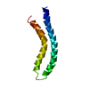
 PDBj
PDBj

 Assembly
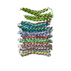
 Sample preparation
 Processing