 Movie
Movie Controller

[English] 日本語
 Yorodumi
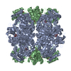
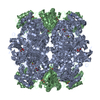
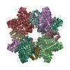
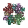
- PDB-1bxn: THE CRYSTAL STRUCTURE OF RUBISCO FROM ALCALIGENES EUTROPHUS TO 2.... -
+ Open data

- Basic information
| Entry | Database: PDB / ID: 1bxn | ||||||
|---|---|---|---|---|---|---|---|
| Title | THE CRYSTAL STRUCTURE OF RUBISCO FROM ALCALIGENES EUTROPHUS TO 2.7 ANGSTROMS. | ||||||
 Components Components |
| ||||||
 Keywords Keywords | LYASE / LYASE (CARBON-CARBON) | ||||||
| Function / homology |  Function and homology information Function and homology informationribulose-bisphosphate carboxylase / ribulose-bisphosphate carboxylase activity / reductive pentose-phosphate cycle / monooxygenase activity / magnesium ion binding Similarity search - Function | ||||||
| Biological species |  Cupriavidus necator (bacteria) Cupriavidus necator (bacteria) | ||||||
| Method |  X-RAY DIFFRACTION / SYNCHROTRON / MOLECULAR REPLACEMENT / Resolution: 2.7 Å X-RAY DIFFRACTION / SYNCHROTRON / MOLECULAR REPLACEMENT / Resolution: 2.7 Å | ||||||
 Authors Authors | Hansen, S. / Vollan, V.B. / Hough, E. / Andersen, K. | ||||||
 Citation Citation | Journal: J.Mol.Biol. / Year: 1999 Title: The crystal structure of rubisco from Alcaligenes eutrophus reveals a novel central eight-stranded beta-barrel formed by beta-strands from four subunits. Authors: Hansen, S. / Vollan, V.B. / Hough, E. / Andersen, K. #1: Journal: Acta Crystallogr.,Sect.D / Year: 1999Title: Purification, Crystallization and Preliminary X-Ray Studies of Two Isoforms of Rubisco from Alcaligenes Eutrophus Authors: Hansen, S. / Hough, E. / Andersen, K. | ||||||
| History |
|
- Structure visualization
| Structure viewer | Molecule: MolmilJmol/JSmol |
|---|
- Downloads & links
-Download
| PDBx/mmCIF format | 1bxn.cif.gz | 405.5 KB | Display | PDBx/mmCIF format |
|---|---|---|---|---|
| PDB format | pdb1bxn.ent.gz | 335.6 KB | Display | PDB format |
| PDBx/mmJSON format | 1bxn.json.gz | Tree view | PDBx/mmJSON format | |
| Others |  Other downloads Other downloads |
-Validation report
| Arichive directory | https://data.pdbj.org/pub/pdb/validation_reports/bx/1bxnftp://data.pdbj.org/pub/pdb/validation_reports/bx/1bxn | HTTPS FTP |
|---|
-Related structure data
| Related structure data |  1ausS S: Starting model for refinement |
|---|---|
| Similar structure data |


-Links
 PDBj
PDBj
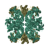
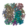
- Assembly
| Deposited unit | 
| ||||||||||||||||||||||||||||
|---|---|---|---|---|---|---|---|---|---|---|---|---|---|---|---|---|---|---|---|---|---|---|---|---|---|---|---|---|---|
| 1 | 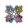
| ||||||||||||||||||||||||||||
| Unit cell |
| ||||||||||||||||||||||||||||
| Noncrystallographic symmetry (NCS) | NCS oper:
|
-Components
| #1: Protein | Mass: 53703.910 Da / Num. of mol.: 4 Source method: isolated from a genetically manipulated source Source: (gene. exp.) Cupriavidus necator (bacteria)References: UniProt: P0C2C2*PLUS, ribulose-bisphosphate carboxylase #2: Protein | Mass: 16058.934 Da / Num. of mol.: 4 Source method: isolated from a genetically manipulated source Source: (gene. exp.) Cupriavidus necator (bacteria)References: UniProt: P09658*PLUS, ribulose-bisphosphate carboxylase #3: Chemical | ChemComp-PO4 /   Mass: 94.971 Da / Num. of mol.: 8 / Source method: obtained synthetically / Formula: PO4 Mass: 94.971 Da / Num. of mol.: 8 / Source method: obtained synthetically / Formula: PO4Has protein modification | Y | |
|---|
-Experimental details
-Experiment
| Experiment | Method: X-RAY DIFFRACTION / Number of used crystals: 1 |
|---|
- Sample preparation
| Crystal | Density Matthews: 2.44 Å3/Da / Density % sol: 45.8 % | ||||||||||||||||||||||||
|---|---|---|---|---|---|---|---|---|---|---|---|---|---|---|---|---|---|---|---|---|---|---|---|---|---|
| Crystal grow | pH: 8.4 / Details: pH 8.40 | ||||||||||||||||||||||||
| Crystal grow | *PLUS Temperature: 4 ℃ / pH: 7.5 / Method: vapor diffusion, sitting drop | ||||||||||||||||||||||||
| Components of the solutions | *PLUS
|
-Data collection
| Diffraction | Mean temperature: 100 K |
|---|---|
| Diffraction source | Source: SYNCHROTRON / Site: ESRF  / Beamline: BM1A / Wavelength: 0.9117 / Beamline: BM1A / Wavelength: 0.9117 |
| Detector | Type: MARRESEARCH / Detector: IMAGE PLATE / Date: May 11, 1997 |
| Radiation | Monochromator: YES / Protocol: SINGLE WAVELENGTH / Monochromatic (M) / Laue (L): M / Scattering type: x-ray |
| Radiation wavelength | Wavelength: 0.9117 Å / Relative weight: 1 |
| Reflection | Resolution: 2.7→25 Å / Num. obs: 65785 / % possible obs: 88 % / Observed criterion σ(I): 0 / Redundancy: 4.4 % / Rmerge(I) obs: 0.125 / Net I/σ(I): 1.5 |
| Reflection | *PLUS % possible obs: 87.5 % |
| Reflection shell | *PLUS Highest resolution: 2.65 Å / Lowest resolution: 2.79 Å / % possible obs: 89.1 % / Redundancy: 4.3 % / Num. unique obs: 9546 / Rmerge(I) obs: 0.695 |
- Processing
| Software |
| ||||||||||||||||||||||||||||||||||||||||||||||||||||||||||||
|---|---|---|---|---|---|---|---|---|---|---|---|---|---|---|---|---|---|---|---|---|---|---|---|---|---|---|---|---|---|---|---|---|---|---|---|---|---|---|---|---|---|---|---|---|---|---|---|---|---|---|---|---|---|---|---|---|---|---|---|---|---|
| Refinement | Method to determine structure: MOLECULAR REPLACEMENT Starting model: 1AUS Resolution: 2.7→15 Å / Cross valid method: THROUGHOUT / σ(F): 1
| ||||||||||||||||||||||||||||||||||||||||||||||||||||||||||||
| Refinement step | Cycle: LAST / Resolution: 2.7→15 Å
| ||||||||||||||||||||||||||||||||||||||||||||||||||||||||||||
| Refine LS restraints |
| ||||||||||||||||||||||||||||||||||||||||||||||||||||||||||||
| Software | *PLUS Name: X-PLOR / Version: 3.8 / Classification: refinement | ||||||||||||||||||||||||||||||||||||||||||||||||||||||||||||
| Refine LS restraints | *PLUS
|
