 Movie
Movie Controller

[English] 日本語
 Yorodumi
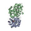
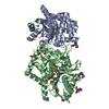
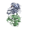
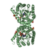
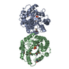
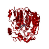
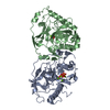
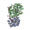
- PDB-1bwd: INOSAMINE-PHOSPHATE AMIDINOTRANSFERASE STRB1 FROM STREPTOMYCES GRISEUS -
+ Open data

- Basic information
| Entry | Database: PDB / ID: 1bwd | ||||||
|---|---|---|---|---|---|---|---|
| Title | INOSAMINE-PHOSPHATE AMIDINOTRANSFERASE STRB1 FROM STREPTOMYCES GRISEUS | ||||||
 Components Components | PROTEIN (INOSAMINE-PHOSPHATE AMIDINOTRANSFERASE) | ||||||
 Keywords Keywords | TRANSFERASE / AMIDINOTRANSFERASE / STREPTOMYCIN | ||||||
| Function / homology |  Function and homology information Function and homology informationscyllo-inosamine-4-phosphate amidinotransferase / scyllo-inosamine-4-phosphate amidinotransferase activity / streptomycin biosynthetic process / glycine amidinotransferase activity / creatine biosynthetic process Similarity search - Function | ||||||
| Biological species |  Streptomyces griseus (bacteria) Streptomyces griseus (bacteria) | ||||||
| Method |  X-RAY DIFFRACTION / MOLECULAR REPLACEMENT / Resolution: 3.1 Å X-RAY DIFFRACTION / MOLECULAR REPLACEMENT / Resolution: 3.1 Å | ||||||
 Authors Authors | Fritsche, E. / Bergner, A. / Humm, A. / Piepersberg, W. / Huber, R. | ||||||
 Citation Citation | Journal: Biochemistry / Year: 1998 Title: Crystal structure of L-arginine:inosamine-phosphate amidinotransferase StrB1 from Streptomyces griseus: an enzyme involved in streptomycin biosynthesis. Authors: Fritsche, E. / Bergner, A. / Humm, A. / Piepersberg, W. / Huber, R. | ||||||
| History |
|
- Structure visualization
| Structure viewer | Molecule: MolmilJmol/JSmol |
|---|
- Downloads & links
-Download
| PDBx/mmCIF format | 1bwd.cif.gz | 142.2 KB | Display | PDBx/mmCIF format |
|---|---|---|---|---|
| PDB format | pdb1bwd.ent.gz | 113 KB | Display | PDB format |
| PDBx/mmJSON format | 1bwd.json.gz | Tree view | PDBx/mmJSON format | |
| Others |  Other downloads Other downloads |
-Validation report
| Summary document | 1bwd_validation.pdf.gz | 378.6 KB | Display | wwPDB validaton report |
|---|---|---|---|---|
| Full document | 1bwd_full_validation.pdf.gz | 409.9 KB | Display | |
| Data in XML | 1bwd_validation.xml.gz | 17.5 KB | Display | |
| Data in CIF | 1bwd_validation.cif.gz | 25.6 KB | Display | |
| Arichive directory | https://data.pdbj.org/pub/pdb/validation_reports/bw/1bwdftp://data.pdbj.org/pub/pdb/validation_reports/bw/1bwd | HTTPS FTP |
-Related structure data
| Related structure data | 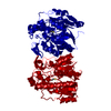 1jdwS S: Starting model for refinement |
|---|---|
| Similar structure data |


-Links
 PDBj
PDBj
- Assembly
| Deposited unit | 
| ||||||||
|---|---|---|---|---|---|---|---|---|---|
| 1 | 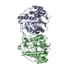
| ||||||||
| Unit cell |
| ||||||||
| Noncrystallographic symmetry (NCS) | NCS oper: (Code: given Matrix: (0.850824, 0.453596, -0.265231), Vector: |
-Components
| #1: Protein | Mass: 38738.645 Da / Num. of mol.: 2 Source method: isolated from a genetically manipulated source Source: (gene. exp.) Streptomyces griseus (bacteria) / Gene: STRB1 / Plasmid: PRSET-STRB1 / Gene (production host): STRB1 / Production host: References: UniProt: P08078, scyllo-inosamine-4-phosphate amidinotransferase |
|---|
-Experimental details
-Experiment
| Experiment | Method: X-RAY DIFFRACTION / Number of used crystals: 1 |
|---|
- Sample preparation
| Crystal | Density Matthews: 2.92 Å3/Da / Density % sol: 58 % | ||||||||||||||||||||||||||||||||||||||||
|---|---|---|---|---|---|---|---|---|---|---|---|---|---|---|---|---|---|---|---|---|---|---|---|---|---|---|---|---|---|---|---|---|---|---|---|---|---|---|---|---|---|
| Crystal grow | pH: 5 Details: 20% PEG 4000, 0.1 M SODIUM CITRAT BUFFER (PH 5.0), 0.2 M AMMONIUM ACETATE | ||||||||||||||||||||||||||||||||||||||||
| Crystal grow | *PLUS Temperature: 20 ℃ / Method: vapor diffusion, hanging dropDetails: drop consists of equal volume of protein and reservoir solutions | ||||||||||||||||||||||||||||||||||||||||
| Components of the solutions | *PLUS
|
-Data collection
| Diffraction | Mean temperature: 283 K |
|---|---|
| Diffraction source | Source: ROTATING ANODE / Type: RIGAKU RU200 / Wavelength: 1.5418 |
| Detector | Type: MARRESEARCH / Detector: IMAGE PLATE |
| Radiation | Monochromator: NI FILTER / Protocol: SINGLE WAVELENGTH / Monochromatic (M) / Laue (L): M / Scattering type: x-ray |
| Radiation wavelength | Wavelength: 1.5418 Å / Relative weight: 1 |
| Reflection | Resolution: 3.1→20 Å / Num. obs: 14273 / % possible obs: 85.6 % / Observed criterion σ(I): 2 / Redundancy: 2.6 % / Rsym value: 0.144 |
| Reflection shell | Resolution: 3.1→3.21 Å / Rsym value: 0.345 / % possible all: 75.3 |
| Reflection | *PLUS Num. measured all: 37728 / Rmerge(I) obs: 0.144 |
| Reflection shell | *PLUS % possible obs: 75.3 % / Rmerge(I) obs: 0.345 |
- Processing
| Software |
| |||||||||||||||||||||||||||||||||||||||||||||||||||||||||||||||
|---|---|---|---|---|---|---|---|---|---|---|---|---|---|---|---|---|---|---|---|---|---|---|---|---|---|---|---|---|---|---|---|---|---|---|---|---|---|---|---|---|---|---|---|---|---|---|---|---|---|---|---|---|---|---|---|---|---|---|---|---|---|---|---|---|
| Refinement | Method to determine structure: MOLECULAR REPLACEMENT Starting model: PDB ID 1JDW Resolution: 3.1→10 Å / σ(F): 0
| |||||||||||||||||||||||||||||||||||||||||||||||||||||||||||||||
| Refinement step | Cycle: LAST / Resolution: 3.1→10 Å
| |||||||||||||||||||||||||||||||||||||||||||||||||||||||||||||||
| Refine LS restraints |
| |||||||||||||||||||||||||||||||||||||||||||||||||||||||||||||||
| Software | *PLUS Name: REFMAC / Classification: refinement | |||||||||||||||||||||||||||||||||||||||||||||||||||||||||||||||
| Refine LS restraints | *PLUS
|
