 Movie
Movie Controller


+ Open data

- Basic information
| Entry | Database: PDB / ID: 1bvp | ||||||
|---|---|---|---|---|---|---|---|
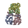
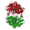
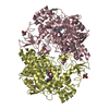
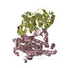
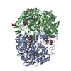
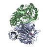
| Title | THE CRYSTAL STRUCTURE OF BLUETONGUE VIRUS VP7 | ||||||
 Components Components | BLUETONGUE VIRUS COAT PROTEIN VP7 | ||||||
 Keywords Keywords | VIRAL PROTEIN / VIRUS / VP7 / TRIMER | ||||||
| Function / homology |  Function and homology information Function and homology informationviral outer capsid / host cell surface receptor binding / fusion of virus membrane with host plasma membrane / viral envelope / structural molecule activity Similarity search - Function | ||||||
| Biological species |  Bluetongue virus Bluetongue virus | ||||||
| Method |  X-RAY DIFFRACTION / Resolution: 2.6 Å X-RAY DIFFRACTION / Resolution: 2.6 Å | ||||||
 Authors Authors | Stuart, D. / Grimes, J. | ||||||
 Citation Citation | Journal: Nature / Year: 1995 Title: The crystal structure of bluetongue virus VP7. Authors: Grimes, J. / Basak, A.K. / Roy, P. / Stuart, D. #1: Journal: J.Mol.Biol. / Year: 1992Title: Preliminary Crystallographic Study Bluetongue Virus Capsid Protein, Vp7 Authors: Basak, A.K. / Stuart, D. / Roy, P. | ||||||
| History |
|
- Structure visualization
| Structure viewer | Molecule: MolmilJmol/JSmol |
|---|
- Downloads & links
-Download
| PDBx/mmCIF format | 1bvp.cif.gz | 390 KB | Display | PDBx/mmCIF format |
|---|---|---|---|---|
| PDB format | pdb1bvp.ent.gz | 324.2 KB | Display | PDB format |
| PDBx/mmJSON format | 1bvp.json.gz | Tree view | PDBx/mmJSON format | |
| Others |  Other downloads Other downloads |
-Validation report
| Arichive directory | https://data.pdbj.org/pub/pdb/validation_reports/bv/1bvpftp://data.pdbj.org/pub/pdb/validation_reports/bv/1bvp | HTTPS FTP |
|---|
-Related structure data
| Similar structure data |
|---|


-Links
 PDBj
PDBj


- Assembly
| Deposited unit | 
| ||||||||||||||||||||||||
|---|---|---|---|---|---|---|---|---|---|---|---|---|---|---|---|---|---|---|---|---|---|---|---|---|---|
| 1 | 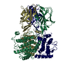
| ||||||||||||||||||||||||
| 2 | 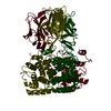
| ||||||||||||||||||||||||
| 3 | 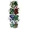
| ||||||||||||||||||||||||
| Unit cell |
| ||||||||||||||||||||||||
| Atom site foot note | 1: CIS PROLINE - PRO 1 338 / 2: CIS PROLINE - PRO 2 338 / 3: CIS PROLINE - PRO 3 338 / 4: CIS PROLINE - PRO 4 338 / 5: CIS PROLINE - PRO 5 338 / 6: CIS PROLINE - PRO 6 338 | ||||||||||||||||||||||||
| Noncrystallographic symmetry (NCS) | NCS oper:
| ||||||||||||||||||||||||
| Details | VP7 FORMS A TRIMER IN CRYSTAL, SOLUTION, AND IN THE VIRUS. MTRIX THE TRANSFORMATIONS PRESENTED ON MTRIX RECORDS BELOW DESCRIBE NON-CRYSTALLOGRAPHIC RELATIONSHIPS AMONG THE VARIOUS DOMAINS IN THIS ENTRY. APPLYING THE APPROPRIATE MTRIX TRANSFORMATION TO THE RESIDUES LISTED FIRST WILL YIELD APPROXIMATE COORDINATES FOR THE RESIDUES LISTED SECOND. APPLIED TO TRANSFORMED TO MTRIX RESIDUES RESIDUES RMSD M1 1 1 .. N 349 2 1 .. N 349 0.0005 M2 1 1 .. N 349 3 1 .. N 349 0.0005 M3 1 1 .. N 349 4 1 .. N 349 0.0005 M4 1 1 .. N 349 5 1 .. N 349 0.0005 M5 1 1 .. N 349 6 1 .. N 349 0.0005 |
-Components
| #1: Protein | Mass: 38578.348 Da / Num. of mol.: 6 Source method: isolated from a genetically manipulated source Source: (gene. exp.) Bluetongue virus / Genus: Orbivirus / Strain: (serotype 10 / American isolate) / References: UniProt: P69361 |
|---|
-Experimental details
-Experiment
| Experiment | Method: X-RAY DIFFRACTION |
|---|
- Sample preparation
| Crystal | Density Matthews: 2.51 Å3/Da / Density % sol: 51.08 % | ||||||||||||||||||||
|---|---|---|---|---|---|---|---|---|---|---|---|---|---|---|---|---|---|---|---|---|---|
| Crystal grow | *PLUS pH: 4.6 / Method: vapor diffusion, hanging drop | ||||||||||||||||||||
| Components of the solutions | *PLUS
|
-Data collection
| Diffraction source | Wavelength: 1.54 |
|---|---|
| Detector | Type: MARRESEARCH |
| Radiation | Scattering type: x-ray |
| Radiation wavelength | Wavelength: 1.54 Å / Relative weight: 1 |
| Reflection | Num. obs: 54361 / % possible obs: 81 % / Observed criterion σ(I): 0 / Rmerge(I) obs: 0.105 |
| Reflection | *PLUS Highest resolution: 2.6 Å / Rmerge(I) obs: 0.105 |
- Processing
| Software |
| ||||||||||||||||||||||||||||||||||||||||||||||||||||||||||||
|---|---|---|---|---|---|---|---|---|---|---|---|---|---|---|---|---|---|---|---|---|---|---|---|---|---|---|---|---|---|---|---|---|---|---|---|---|---|---|---|---|---|---|---|---|---|---|---|---|---|---|---|---|---|---|---|---|---|---|---|---|---|
| Refinement | Resolution: 2.6→15 Å / σ(F): 0 /
| ||||||||||||||||||||||||||||||||||||||||||||||||||||||||||||
| Displacement parameters | Biso mean: 23.8 Å2 | ||||||||||||||||||||||||||||||||||||||||||||||||||||||||||||
| Refinement step | Cycle: LAST / Resolution: 2.6→15 Å
| ||||||||||||||||||||||||||||||||||||||||||||||||||||||||||||
| Refine LS restraints |
| ||||||||||||||||||||||||||||||||||||||||||||||||||||||||||||
| Software | *PLUS Name: X-PLOR / Classification: refinement | ||||||||||||||||||||||||||||||||||||||||||||||||||||||||||||
| Refinement | *PLUS | ||||||||||||||||||||||||||||||||||||||||||||||||||||||||||||
| Solvent computation | *PLUS | ||||||||||||||||||||||||||||||||||||||||||||||||||||||||||||
| Displacement parameters | *PLUS | ||||||||||||||||||||||||||||||||||||||||||||||||||||||||||||
| Refine LS restraints | *PLUS Type: x_angle_deg / Dev ideal: 1.85 |
