 Movie
Movie Controller


+ Open data

- Basic information
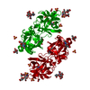
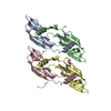
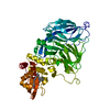
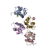
| Entry | Database: PDB / ID: 1bv4 | ||||||
|---|---|---|---|---|---|---|---|
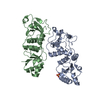
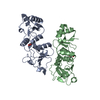
| Title | APO-MANNOSE-BINDING PROTEIN-C | ||||||
 Components Components | PROTEIN (MANNOSE-BINDING PROTEIN-C) | ||||||
 Keywords Keywords | SUGAR BINDING PROTEIN / C-TYPE LECTIN / CALCIUM-BINDING PROTEIN / COLLECTIN | ||||||
| Function / homology |  Function and homology information Function and homology informationLectin pathway of complement activation / positive regulation of opsonization / positive regulation of protein processing / positive regulation of complement activation / complement activation, lectin pathway / negative regulation of viral process / galactose binding / : / cell surface pattern recognition receptor signaling pathway / serine-type endopeptidase complex ...Lectin pathway of complement activation / positive regulation of opsonization / positive regulation of protein processing / positive regulation of complement activation / complement activation, lectin pathway / negative regulation of viral process / galactose binding / : / cell surface pattern recognition receptor signaling pathway / serine-type endopeptidase complex / surfactant homeostasis / symbiont cell surface / phosphatidylinositol-4-phosphate binding / pattern recognition receptor activity / D-mannose binding / complement activation, classical pathway / multivesicular body / positive regulation of phagocytosis / antiviral innate immune response / calcium-dependent protein binding / protease binding / defense response to Gram-positive bacterium / signaling receptor binding / innate immune response / calcium ion binding / cell surface / protein-containing complex / proteolysis / : / identical protein binding Similarity search - Function | ||||||
| Biological species |  | ||||||
| Method |  X-RAY DIFFRACTION / SYNCHROTRON / MOLECULAR REPLACEMENT / Resolution: 1.85 Å X-RAY DIFFRACTION / SYNCHROTRON / MOLECULAR REPLACEMENT / Resolution: 1.85 Å | ||||||
 Authors Authors | Ng, K.K.-S. / Weis, W.I. | ||||||
 Citation Citation | Journal: Biochemistry / Year: 1998 Title: Ca2+-dependent structural changes in C-type mannose-binding proteins. Authors: Ng, K.K. / Park-Snyder, S. / Weis, W.I. | ||||||
| History |
|
- Structure visualization
| Structure viewer | Molecule: MolmilJmol/JSmol |
|---|
- Downloads & links
-Download
| PDBx/mmCIF format | 1bv4.cif.gz | 95 KB | Display | PDBx/mmCIF format |
|---|---|---|---|---|
| PDB format | pdb1bv4.ent.gz | 73.4 KB | Display | PDB format |
| PDBx/mmJSON format | 1bv4.json.gz | Tree view | PDBx/mmJSON format | |
| Others |  Other downloads Other downloads |
-Validation report
| Arichive directory | https://data.pdbj.org/pub/pdb/validation_reports/bv/1bv4ftp://data.pdbj.org/pub/pdb/validation_reports/bv/1bv4 | HTTPS FTP |
|---|
-Related structure data
| Related structure data |  1buuC  1rdoS S: Starting model for refinement C: citing same article ( |
|---|---|
| Similar structure data |


-Links
 PDBj
PDBj


- Assembly
| Deposited unit | 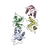
| ||||||||||||||||
|---|---|---|---|---|---|---|---|---|---|---|---|---|---|---|---|---|---|
| 1 |
| ||||||||||||||||
| Unit cell |
| ||||||||||||||||
| Noncrystallographic symmetry (NCS) | NCS oper:
|
-Components
| #1: Protein | Mass: 13161.604 Da / Num. of mol.: 4 / Fragment: SUBTILISIN FRAGMENT Source method: isolated from a genetically manipulated source Source: (gene. exp.)  #2: Water | ChemComp-HOH / |  Mass: 18.015 Da / Num. of mol.: 234 / Source method: isolated from a natural source / Formula: H2O Mass: 18.015 Da / Num. of mol.: 234 / Source method: isolated from a natural source / Formula: H2OHas protein modification | Y | |
|---|
-Experimental details
-Experiment
| Experiment | Method: X-RAY DIFFRACTION / Number of used crystals: 1 |
|---|
- Sample preparation
| Crystal | Density Matthews: 2.18 Å3/Da / Density % sol: 36 % | |||||||||||||||||||||||||||||||||||||||||||||||||
|---|---|---|---|---|---|---|---|---|---|---|---|---|---|---|---|---|---|---|---|---|---|---|---|---|---|---|---|---|---|---|---|---|---|---|---|---|---|---|---|---|---|---|---|---|---|---|---|---|---|---|
| Crystal grow | pH: 6.2 Details: 12% PEG 8000, 100 MM NAMES, PH 6.1, 200 MM LICL, 2 MM EDTA 0.02%, NAN3, pH 6.2 | |||||||||||||||||||||||||||||||||||||||||||||||||
| Crystal | *PLUS | |||||||||||||||||||||||||||||||||||||||||||||||||
| Crystal grow | *PLUS Method: vapor diffusion, hanging drop / pH: 8 | |||||||||||||||||||||||||||||||||||||||||||||||||
| Components of the solutions | *PLUS
|
-Data collection
| Diffraction | Mean temperature: 100 K |
|---|---|
| Diffraction source | Source: SYNCHROTRON / Site: SSRL  / Beamline: BL7-1 / Wavelength: 1.08 / Beamline: BL7-1 / Wavelength: 1.08 |
| Detector | Type: MARRESEARCH / Detector: IMAGE PLATE / Date: May 15, 1996 / Details: MIRRORS |
| Radiation | Protocol: SINGLE WAVELENGTH / Monochromatic (M) / Laue (L): M / Scattering type: x-ray |
| Radiation wavelength | Wavelength: 1.08 Å / Relative weight: 1 |
| Reflection | Resolution: 1.85→50 Å / Num. obs: 33667 / % possible obs: 99.9 % / Observed criterion σ(I): 0 / Redundancy: 3.7 % / Biso Wilson estimate: 15.6 Å2 / Rmerge(I) obs: 0.039 / Net I/σ(I): 20.6 |
| Reflection shell | Resolution: 1.85→1.92 Å / Redundancy: 3.5 % / Rmerge(I) obs: 0.26 / Mean I/σ(I) obs: 6.2 / % possible all: 99.8 |
| Reflection shell | *PLUS % possible obs: 99.8 % / Rmerge(I) obs: 0.26 |
- Processing
| Software |
| ||||||||||||||||||||||||||||||||||||||||||||||||||||||||||||||||||||||||||||||||
|---|---|---|---|---|---|---|---|---|---|---|---|---|---|---|---|---|---|---|---|---|---|---|---|---|---|---|---|---|---|---|---|---|---|---|---|---|---|---|---|---|---|---|---|---|---|---|---|---|---|---|---|---|---|---|---|---|---|---|---|---|---|---|---|---|---|---|---|---|---|---|---|---|---|---|---|---|---|---|---|---|---|
| Refinement | Method to determine structure: MOLECULAR REPLACEMENT Starting model: 1RDO Resolution: 1.85→50 Å / Rfactor Rfree error: 0.004 / Data cutoff high rms absF: 10000 / Isotropic thermal model: RESTRAINED / Cross valid method: THROUGHOUT / σ(F): 0
| ||||||||||||||||||||||||||||||||||||||||||||||||||||||||||||||||||||||||||||||||
| Solvent computation | Solvent model: FLAT MODEL / Bsol: 38.1 Å2 / ksol: 0.32 e/Å3 | ||||||||||||||||||||||||||||||||||||||||||||||||||||||||||||||||||||||||||||||||
| Displacement parameters | Biso mean: 27.3 Å2
| ||||||||||||||||||||||||||||||||||||||||||||||||||||||||||||||||||||||||||||||||
| Refine analyze |
| ||||||||||||||||||||||||||||||||||||||||||||||||||||||||||||||||||||||||||||||||
| Refinement step | Cycle: LAST / Resolution: 1.85→50 Å
| ||||||||||||||||||||||||||||||||||||||||||||||||||||||||||||||||||||||||||||||||
| Refine LS restraints |
| ||||||||||||||||||||||||||||||||||||||||||||||||||||||||||||||||||||||||||||||||
| LS refinement shell | Resolution: 1.85→1.92 Å / Rfactor Rfree error: 0.018 / Total num. of bins used: 10
| ||||||||||||||||||||||||||||||||||||||||||||||||||||||||||||||||||||||||||||||||
| Xplor file |
| ||||||||||||||||||||||||||||||||||||||||||||||||||||||||||||||||||||||||||||||||
| Software | *PLUS Name: CNS / Version: 0.3C / Classification: refinement | ||||||||||||||||||||||||||||||||||||||||||||||||||||||||||||||||||||||||||||||||
| Refinement | *PLUS Lowest resolution: 50 Å / σ(F): 0 / % reflection Rfree: 10 % | ||||||||||||||||||||||||||||||||||||||||||||||||||||||||||||||||||||||||||||||||
| Solvent computation | *PLUS | ||||||||||||||||||||||||||||||||||||||||||||||||||||||||||||||||||||||||||||||||
| Displacement parameters | *PLUS Biso mean: 27.3 Å2 | ||||||||||||||||||||||||||||||||||||||||||||||||||||||||||||||||||||||||||||||||
| Refine LS restraints | *PLUS
| ||||||||||||||||||||||||||||||||||||||||||||||||||||||||||||||||||||||||||||||||
| LS refinement shell | *PLUS Rfactor Rfree: 0.304 / % reflection Rfree: 10 % / Rfactor Rwork: 0.237 / Rfactor obs: 0.237 |
