 Movie
Movie Controller


+ Open data

- Basic information
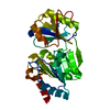
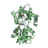
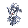
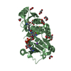
| Entry | Database: PDB / ID: 1bu2 | ||||||
|---|---|---|---|---|---|---|---|
| Title | X-RAY STRUCTURE OF A VIRAL CYCLIN FROM HERPESVIRUS SAIMIRI | ||||||
 Components Components | CYCLIN HOMOLOG | ||||||
 Keywords Keywords | CELL CYCLE REGULATION / HERPESVIRUS SAIMIRI / VIRAL CYCLIN | ||||||
| Function / homology |  Function and homology information Function and homology informationsymbiont-mediated perturbation of host cell cycle progression / cell division Similarity search - Function | ||||||
| Biological species | Herpesvirus saimiri | ||||||
| Method |  X-RAY DIFFRACTION / SYNCHROTRON / MOLECULAR REPLACEMENT / Resolution: 3 Å X-RAY DIFFRACTION / SYNCHROTRON / MOLECULAR REPLACEMENT / Resolution: 3 Å | ||||||
 Authors Authors | Schulze-Gahmen, U. / Jung, J.U. / Kim, S.-H. | ||||||
 Citation Citation | Journal: Structure Fold.Des. / Year: 1999 Title: Crystal structure of a viral cyclin, a positive regulator of cyclin-dependent kinase 6. Authors: Schulze-Gahmen, U. / Jung, J.U. / Kim, S.H. #1: Journal: Mol.Cell.Biol. / Year: 1994Title: Virus-Encoded Cyclin Authors: Jung, J.U. / Staeger, M. / Desrosier, R.C. | ||||||
| History |
|
- Structure visualization
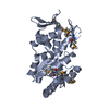
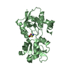
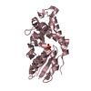
| Structure viewer | Molecule: MolmilJmol/JSmol |
|---|
- Downloads & links
-Download
| PDBx/mmCIF format | 1bu2.cif.gz | 50.6 KB | Display | PDBx/mmCIF format |
|---|---|---|---|---|
| PDB format | pdb1bu2.ent.gz | 36.1 KB | Display | PDB format |
| PDBx/mmJSON format | 1bu2.json.gz | Tree view | PDBx/mmJSON format | |
| Others |  Other downloads Other downloads |
-Validation report
| Arichive directory | https://data.pdbj.org/pub/pdb/validation_reports/bu/1bu2ftp://data.pdbj.org/pub/pdb/validation_reports/bu/1bu2 | HTTPS FTP |
|---|
-Related structure data
| Similar structure data |
|---|


-Links
 PDBj
PDBj


- Assembly
| Deposited unit | 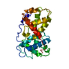
| ||||||||
|---|---|---|---|---|---|---|---|---|---|
| 1 |
| ||||||||
| Unit cell |
|
-Components
| #1: Protein | Mass: 25849.154 Da / Num. of mol.: 1 Source method: isolated from a genetically manipulated source Source: (gene. exp.)  Herpesvirus saimiri (strain 11) / Genus: Rhadinovirus / Species: Saimiriine herpesvirus 2 / Strain: 11 / Gene: ECLF2 / Plasmid: PQE9 / Production host: Herpesvirus saimiri (strain 11) / Genus: Rhadinovirus / Species: Saimiriine herpesvirus 2 / Strain: 11 / Gene: ECLF2 / Plasmid: PQE9 / Production host:  |
|---|
-Experimental details
-Experiment
| Experiment | Method: X-RAY DIFFRACTION / Number of used crystals: 1 |
|---|
- Sample preparation
| Crystal | Density Matthews: 2.94 Å3/Da / Density % sol: 58.18 % | |||||||||||||||||||||||||||||||||||
|---|---|---|---|---|---|---|---|---|---|---|---|---|---|---|---|---|---|---|---|---|---|---|---|---|---|---|---|---|---|---|---|---|---|---|---|---|
| Crystal grow | pH: 7 / Details: pH 7.0 | |||||||||||||||||||||||||||||||||||
| Crystal grow | *PLUS Temperature: 22 ℃ / Method: vapor diffusion, sitting drop | |||||||||||||||||||||||||||||||||||
| Components of the solutions | *PLUS
|
-Data collection
| Diffraction | Mean temperature: 100 K |
|---|---|
| Diffraction source | Source: SYNCHROTRON / Site: NSLS  / Beamline: X12B / Wavelength: 0.9471 / Beamline: X12B / Wavelength: 0.9471 |
| Detector | Type: MARRESEARCH / Date: Nov 1, 1995 |
| Radiation | Monochromatic (M) / Laue (L): M / Scattering type: x-ray |
| Radiation wavelength | Wavelength: 0.9471 Å / Relative weight: 1 |
| Reflection | Resolution: 3→20 Å / Num. obs: 6501 / % possible obs: 98.9 % / Redundancy: 4 % / Biso Wilson estimate: 69.2 Å2 / Rsym value: 0.95 / Net I/σ(I): 11.5 |
| Reflection | *PLUS Num. measured all: 23083 / Rmerge(I) obs: 0.095 |
| Reflection shell | *PLUS % possible obs: 99.8 % / Rmerge(I) obs: 0.35 / Mean I/σ(I) obs: 2.8 |
- Processing
| Software |
| ||||||||||||||||||||||||||||||||||||||||||||||||||||||||||||
|---|---|---|---|---|---|---|---|---|---|---|---|---|---|---|---|---|---|---|---|---|---|---|---|---|---|---|---|---|---|---|---|---|---|---|---|---|---|---|---|---|---|---|---|---|---|---|---|---|---|---|---|---|---|---|---|---|---|---|---|---|---|
| Refinement | Method to determine structure: MOLECULAR REPLACEMENT / Resolution: 3→20 Å / Rfactor Rfree error: 0.012 / Data cutoff high rms absF: 1077669.13 / Isotropic thermal model: GROUP / Cross valid method: THROUGHOUT / σ(F): 2 / Details: BULK SOLVENT MODEL USED
| ||||||||||||||||||||||||||||||||||||||||||||||||||||||||||||
| Displacement parameters | Biso mean: 64.7 Å2
| ||||||||||||||||||||||||||||||||||||||||||||||||||||||||||||
| Refine analyze |
| ||||||||||||||||||||||||||||||||||||||||||||||||||||||||||||
| Refinement step | Cycle: LAST / Resolution: 3→20 Å
| ||||||||||||||||||||||||||||||||||||||||||||||||||||||||||||
| Refine LS restraints |
| ||||||||||||||||||||||||||||||||||||||||||||||||||||||||||||
| LS refinement shell | Resolution: 3→3.19 Å / Rfactor Rfree error: 0.044 / Total num. of bins used: 6
| ||||||||||||||||||||||||||||||||||||||||||||||||||||||||||||
| Xplor file |
| ||||||||||||||||||||||||||||||||||||||||||||||||||||||||||||
| Software | *PLUS Name: CNS / Version: 0.3 / Classification: refinement | ||||||||||||||||||||||||||||||||||||||||||||||||||||||||||||
| Refinement | *PLUS Rfactor Rfree: 0.324 | ||||||||||||||||||||||||||||||||||||||||||||||||||||||||||||
| Solvent computation | *PLUS | ||||||||||||||||||||||||||||||||||||||||||||||||||||||||||||
| Displacement parameters | *PLUS | ||||||||||||||||||||||||||||||||||||||||||||||||||||||||||||
| Refine LS restraints | *PLUS
|
