 Movie
Movie Controller

[English] 日本語
 Yorodumi
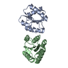
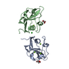
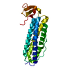
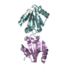
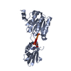
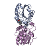
- PDB-1btn: STRUCTURE OF THE BINDING SITE FOR INOSITOL PHOSPHATES IN A PH DOMAIN -
+ Open data

- Basic information
| Entry | Database: PDB / ID: 1btn | ||||||
|---|---|---|---|---|---|---|---|
| Title | STRUCTURE OF THE BINDING SITE FOR INOSITOL PHOSPHATES IN A PH DOMAIN | ||||||
 Components Components | BETA-SPECTRIN | ||||||
 Keywords Keywords | SIGNAL TRANSDUCTION PROTEIN | ||||||
| Function / homology |  Function and homology information Function and homology informationInteraction between L1 and Ankyrins / regulation of SMAD protein signal transduction / RHOV GTPase cycle / NCAM signaling for neurite out-growth / membrane assembly / central nervous system formation / RHOU GTPase cycle / spectrin / cuticular plate / COPI-mediated anterograde transport ...Interaction between L1 and Ankyrins / regulation of SMAD protein signal transduction / RHOV GTPase cycle / NCAM signaling for neurite out-growth / membrane assembly / central nervous system formation / RHOU GTPase cycle / spectrin / cuticular plate / COPI-mediated anterograde transport / plasma membrane organization / RAF/MAP kinase cascade / Golgi to plasma membrane protein transport / actin filament capping / M band / ankyrin binding / cortical actin cytoskeleton / cortical cytoskeleton / mitotic cytokinesis / axolemma / positive regulation of interleukin-2 production / regulation of protein localization to plasma membrane / cell projection / endomembrane system / protein localization to plasma membrane / positive regulation of protein localization to plasma membrane / central nervous system development / phospholipid binding / structural constituent of cytoskeleton / actin filament binding / cell junction / actin cytoskeleton organization / GTPase binding / calmodulin binding / postsynaptic density / postsynapse / protein-containing complex binding / nucleolus / glutamatergic synapse / protein-containing complex / membrane / nucleus / plasma membrane / cytoplasm / cytosol Similarity search - Function | ||||||
| Biological species |  | ||||||
| Method |  X-RAY DIFFRACTION / Resolution: 2 Å X-RAY DIFFRACTION / Resolution: 2 Å | ||||||
 Authors Authors | Wilmanns, M. / Hyvoenen, M. / Saraste, M. | ||||||
 Citation Citation | Journal: EMBO J. / Year: 1995 Title: Structure of the binding site for inositol phosphates in a PH domain. Authors: Hyvonen, M. / Macias, M.J. / Nilges, M. / Oschkinat, H. / Saraste, M. / Wilmanns, M. #1: Journal: Nature / Year: 1994Title: Structure of the Ph Domain from Beta-Spectrin Authors: Macias, M.J. / Musacchio, A. / Postingl, H. / Nilges, M. / Saraste, M. / Oschkinat, H. | ||||||
| History |
|
- Structure visualization
| Structure viewer | Molecule: MolmilJmol/JSmol |
|---|
- Downloads & links
-Download
| PDBx/mmCIF format | 1btn.cif.gz | 37.1 KB | Display | PDBx/mmCIF format |
|---|---|---|---|---|
| PDB format | pdb1btn.ent.gz | 23.8 KB | Display | PDB format |
| PDBx/mmJSON format | 1btn.json.gz | Tree view | PDBx/mmJSON format | |
| Others |  Other downloads Other downloads |
-Validation report
| Arichive directory | https://data.pdbj.org/pub/pdb/validation_reports/bt/1btnftp://data.pdbj.org/pub/pdb/validation_reports/bt/1btn | HTTPS FTP |
|---|
-Related structure data
| Similar structure data |
|---|


-Links
 PDBj
PDBj

- Assembly
| Deposited unit | 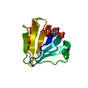
| ||||||||
|---|---|---|---|---|---|---|---|---|---|
| 1 | 
| ||||||||
| Unit cell |
|
-Components
| #1: Protein | Mass: 12287.853 Da / Num. of mol.: 1 / Fragment: PH DOMAIN, RESIDUES 2199 - 2304 Source method: isolated from a genetically manipulated source Source: (gene. exp.)  |
|---|---|
| #2: Chemical | ChemComp-I3P / 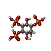  Mass: 420.096 Da / Num. of mol.: 1 / Source method: obtained synthetically / Formula: C6H15O15P3 Mass: 420.096 Da / Num. of mol.: 1 / Source method: obtained synthetically / Formula: C6H15O15P3 |
| #3: Water | ChemComp-HOH /  Mass: 18.015 Da / Num. of mol.: 77 / Source method: isolated from a natural source / Formula: H2O Mass: 18.015 Da / Num. of mol.: 77 / Source method: isolated from a natural source / Formula: H2O |
-Experimental details
-Experiment
| Experiment | Method: X-RAY DIFFRACTION |
|---|
- Sample preparation
| Crystal | Density Matthews: 2.46 Å3/Da / Density % sol: 50 % | ||||||||||||||||||||||||||||||||||||||||||
|---|---|---|---|---|---|---|---|---|---|---|---|---|---|---|---|---|---|---|---|---|---|---|---|---|---|---|---|---|---|---|---|---|---|---|---|---|---|---|---|---|---|---|---|
| Crystal grow | pH: 6.5 / Details: pH 6.5 | ||||||||||||||||||||||||||||||||||||||||||
| Crystal grow | *PLUS Method: vapor diffusion, hanging drop | ||||||||||||||||||||||||||||||||||||||||||
| Components of the solutions | *PLUS
|
-Data collection
| Diffraction | Mean temperature: 293 K |
|---|---|
| Diffraction source | Wavelength: 1.54 |
| Detector | Type: MARRESEARCH / Detector: IMAGE PLATE / Date: Feb 1, 1995 |
| Radiation | Monochromatic (M) / Laue (L): M / Scattering type: x-ray |
| Radiation wavelength | Wavelength: 1.54 Å / Relative weight: 1 |
| Reflection | Resolution: 2→1000 Å / Num. obs: 7830 / % possible obs: 89.6 % / Observed criterion σ(I): 0 / Redundancy: 5.4 % / Rmerge(I) obs: 0.047 |
| Reflection | *PLUS Num. measured all: 41713 / Rmerge(I) obs: 0.047 |
- Processing
| Software |
| ||||||||||||||||||||||||||||||||||||||||||||||||||||||||||||
|---|---|---|---|---|---|---|---|---|---|---|---|---|---|---|---|---|---|---|---|---|---|---|---|---|---|---|---|---|---|---|---|---|---|---|---|---|---|---|---|---|---|---|---|---|---|---|---|---|---|---|---|---|---|---|---|---|---|---|---|---|---|
| Refinement | Resolution: 2→8 Å / σ(F): 1
| ||||||||||||||||||||||||||||||||||||||||||||||||||||||||||||
| Displacement parameters | Biso mean: 27.9 Å2 | ||||||||||||||||||||||||||||||||||||||||||||||||||||||||||||
| Refine analyze | Luzzati coordinate error obs: 0.27 Å | ||||||||||||||||||||||||||||||||||||||||||||||||||||||||||||
| Refinement step | Cycle: LAST / Resolution: 2→8 Å
| ||||||||||||||||||||||||||||||||||||||||||||||||||||||||||||
| Refine LS restraints |
| ||||||||||||||||||||||||||||||||||||||||||||||||||||||||||||
| Software | *PLUS Name: X-PLOR / Classification: refinement | ||||||||||||||||||||||||||||||||||||||||||||||||||||||||||||
| Refinement | *PLUS | ||||||||||||||||||||||||||||||||||||||||||||||||||||||||||||
| Solvent computation | *PLUS | ||||||||||||||||||||||||||||||||||||||||||||||||||||||||||||
| Displacement parameters | *PLUS | ||||||||||||||||||||||||||||||||||||||||||||||||||||||||||||
| Refine LS restraints | *PLUS
|
