 Movie
Movie Controller


+ Open data

- Basic information
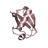
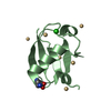
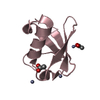
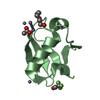
| Entry | Database: PDB / ID: 1bt0 | ||||||
|---|---|---|---|---|---|---|---|
| Title | STRUCTURE OF UBIQUITIN-LIKE PROTEIN, RUB1 | ||||||
 Components Components | PROTEIN (UBIQUITIN-LIKE PROTEIN 7, RUB1) | ||||||
 Keywords Keywords | SIGNALING PROTEIN / RUB1 / UBIQUITIN-LIKE PROTEIN / ARABIDOPSIS | ||||||
| Function / homology |  Function and homology information Function and homology informationethylene biosynthetic process / response to auxin / protein neddylation / cytosolic ribosome / modification-dependent protein catabolic process / protein tag activity / structural constituent of ribosome / protein ubiquitination / mRNA binding / nucleus ...ethylene biosynthetic process / response to auxin / protein neddylation / cytosolic ribosome / modification-dependent protein catabolic process / protein tag activity / structural constituent of ribosome / protein ubiquitination / mRNA binding / nucleus / plasma membrane / cytosol Similarity search - Function | ||||||
| Biological species |  | ||||||
| Method |  X-RAY DIFFRACTION / MOLECULAR REPLACEMENT / Resolution: 1.7 Å X-RAY DIFFRACTION / MOLECULAR REPLACEMENT / Resolution: 1.7 Å | ||||||
 Authors Authors | Delacruz, W.P. / Fisher, A.J. | ||||||
 Citation Citation | Journal: J.Biol.Chem. / Year: 1998 Title: The rub family of ubiquitin-like proteins. Crystal structure of Arabidopsis rub1 and expression of multiple rubs in Arabidopsis. Authors: Rao-Naik, C. / delaCruz, W. / Laplaza, J.M. / Tan, S. / Callis, J. / Fisher, A.J. #1: Journal: J.Mol.Biol. / Year: 1987Title: Structure of Ubiquitin Refined at 1.8 Angstroms Resolution Authors: Vijay-Kumar, S. / Bugg, C.E. / Cook, W.J. | ||||||
| History |
|
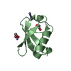
- Structure visualization
| Structure viewer | Molecule: MolmilJmol/JSmol |
|---|
- Downloads & links
-Download
| PDBx/mmCIF format | 1bt0.cif.gz | 29 KB | Display | PDBx/mmCIF format |
|---|---|---|---|---|
| PDB format | pdb1bt0.ent.gz | 18 KB | Display | PDB format |
| PDBx/mmJSON format | 1bt0.json.gz | Tree view | PDBx/mmJSON format | |
| Others |  Other downloads Other downloads |
-Validation report
| Arichive directory | https://data.pdbj.org/pub/pdb/validation_reports/bt/1bt0ftp://data.pdbj.org/pub/pdb/validation_reports/bt/1bt0 | HTTPS FTP |
|---|
-Related structure data
| Related structure data |  1ubiS S: Starting model for refinement |
|---|---|
| Similar structure data |


-Links
 PDBj
PDBj
- Assembly
| Deposited unit | 
| ||||||||
|---|---|---|---|---|---|---|---|---|---|
| 1 |
| ||||||||
| Unit cell |
|
-Components
| #1: Protein | Mass: 8484.797 Da / Num. of mol.: 1 / Mutation: T1M, M2L, A60N Source method: isolated from a genetically manipulated source Source: (gene. exp.)  | ||||
|---|---|---|---|---|---|
| #2: Chemical |   Mass: 65.409 Da / Num. of mol.: 2 / Source method: obtained synthetically / Formula: Zn Mass: 65.409 Da / Num. of mol.: 2 / Source method: obtained synthetically / Formula: Zn#3: Chemical |   Mass: 62.068 Da / Num. of mol.: 3 / Source method: obtained synthetically / Formula: C2H6O2 Mass: 62.068 Da / Num. of mol.: 3 / Source method: obtained synthetically / Formula: C2H6O2#4: Water | ChemComp-HOH / |  Mass: 18.015 Da / Num. of mol.: 75 / Source method: isolated from a natural source / Formula: H2O Mass: 18.015 Da / Num. of mol.: 75 / Source method: isolated from a natural source / Formula: H2O |
-Experimental details
-Experiment
| Experiment | Method: X-RAY DIFFRACTION / Number of used crystals: 1 |
|---|
- Sample preparation
| Crystal | Density Matthews: 2.22 Å3/Da / Density % sol: 46 % | ||||||||||||||||||||||||||||||
|---|---|---|---|---|---|---|---|---|---|---|---|---|---|---|---|---|---|---|---|---|---|---|---|---|---|---|---|---|---|---|---|
| Crystal grow | pH: 6.5 / Details: pH 6.5 | ||||||||||||||||||||||||||||||
| Crystal | *PLUS Density % sol: 46 % | ||||||||||||||||||||||||||||||
| Crystal grow | *PLUS Method: batch method | ||||||||||||||||||||||||||||||
| Components of the solutions | *PLUS
|
-Data collection
| Diffraction | Mean temperature: 100 K |
|---|---|
| Diffraction source | Source: ROTATING ANODE / Type: RIGAKU / Wavelength: 1.5418 |
| Detector | Type: SIEMENS / Detector: AREA DETECTOR / Date: Aug 15, 1997 |
| Radiation | Protocol: SINGLE WAVELENGTH / Monochromatic (M) / Laue (L): M / Scattering type: x-ray |
| Radiation wavelength | Wavelength: 1.5418 Å / Relative weight: 1 |
| Reflection | Resolution: 1.7→30 Å / Num. obs: 7919 / % possible obs: 95 % / Observed criterion σ(I): 0 / Redundancy: 2.9 % / Rmerge(I) obs: 0.051 |
| Reflection | *PLUS Num. measured all: 23228 |
- Processing
| Software |
| ||||||||||||||||||||||||||||||
|---|---|---|---|---|---|---|---|---|---|---|---|---|---|---|---|---|---|---|---|---|---|---|---|---|---|---|---|---|---|---|---|
| Refinement | Method to determine structure: MOLECULAR REPLACEMENT Starting model: PDB ENTRY 1UBI WITH NON-CONSERVED RESIDUES TRUNCATED INTO ALANINE Resolution: 1.7→30 Å / σ(F): 0 / Stereochemistry target values: TNT PROTGEO /
| ||||||||||||||||||||||||||||||
| Solvent computation | Solvent model: TNT / Bsol: 461 Å2 / ksol: 1.018 e/Å3 | ||||||||||||||||||||||||||||||
| Refinement step | Cycle: LAST / Resolution: 1.7→30 Å
| ||||||||||||||||||||||||||||||
| Refine LS restraints |
| ||||||||||||||||||||||||||||||
| Software | *PLUS Name: TNT / Classification: refinement | ||||||||||||||||||||||||||||||
| Refinement | *PLUS σ(F): 0 / % reflection Rfree: 5 % / Rfactor obs: 0.18 / Rfactor Rfree: 0.246 / Rfactor Rwork: 0.18 | ||||||||||||||||||||||||||||||
| Solvent computation | *PLUS | ||||||||||||||||||||||||||||||
| Displacement parameters | *PLUS | ||||||||||||||||||||||||||||||
| Refine LS restraints | *PLUS
|
