 Movie
Movie Controller


+ Open data

- Basic information
| Entry | Database: PDB / ID: 1bs0 | ||||||
|---|---|---|---|---|---|---|---|
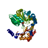
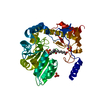
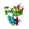
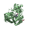
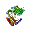
| Title | PLP-DEPENDENT ACYL-COA SYNTHASE | ||||||
 Components Components | PROTEIN (8-AMINO-7-OXONANOATE SYNTHASE) | ||||||
 Keywords Keywords | TRANSFERASE / PLP-DEPENDENT ACYL-COA SYNTHASE / BIOTIN BIOSYNTHESIS / 8-AMINO-7-OXONANOATE SYNTHASE / 8-AMINO-7-KETOPELARGONATE SYNTHASE | ||||||
| Function / homology |  Function and homology information Function and homology information8-amino-7-oxononanoate synthase / 8-amino-7-oxononanoate synthase activity / biotin biosynthetic process / pyridoxal phosphate binding / protein homodimerization activity Similarity search - Function | ||||||
| Biological species |  | ||||||
| Method |  X-RAY DIFFRACTION / SYNCHROTRON / MIR / Resolution: 1.65 Å X-RAY DIFFRACTION / SYNCHROTRON / MIR / Resolution: 1.65 Å | ||||||
 Authors Authors | Alexeev, D. / Alexeeva, M. / Baxter, R.L. / Campopiano, D.J. / Webster, S.P. / Sawyer, L. | ||||||
 Citation Citation | Journal: J.Mol.Biol. / Year: 1998 Title: The crystal structure of 8-amino-7-oxononanoate synthase: a bacterial PLP-dependent, acyl-CoA-condensing enzyme. Authors: Alexeev, D. / Alexeeva, M. / Baxter, R.L. / Campopiano, D.J. / Webster, S.P. / Sawyer, L. | ||||||
| History |
|
- Structure visualization
| Structure viewer | Molecule: MolmilJmol/JSmol |
|---|
- Downloads & links
-Download
| PDBx/mmCIF format | 1bs0.cif.gz | 99.7 KB | Display | PDBx/mmCIF format |
|---|---|---|---|---|
| PDB format | pdb1bs0.ent.gz | 75.8 KB | Display | PDB format |
| PDBx/mmJSON format | 1bs0.json.gz | Tree view | PDBx/mmJSON format | |
| Others |  Other downloads Other downloads |
-Validation report
| Arichive directory | https://data.pdbj.org/pub/pdb/validation_reports/bs/1bs0ftp://data.pdbj.org/pub/pdb/validation_reports/bs/1bs0 | HTTPS FTP |
|---|
-Related structure data
| Similar structure data |
|---|


-Links
 PDBj
PDBj- Assembly
| Deposited unit | 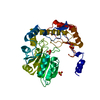
| |||||||||||||||
|---|---|---|---|---|---|---|---|---|---|---|---|---|---|---|---|---|
| 1 |
| |||||||||||||||
| 2 | 
| |||||||||||||||
| Unit cell |
| |||||||||||||||
| Components on special symmetry positions |
|
-Components
| #1: Protein | Mass: 41641.172 Da / Num. of mol.: 1 Source method: isolated from a genetically manipulated source Source: (gene. exp.) References: UniProt: P12998, 8-amino-7-oxononanoate synthase | ||
|---|---|---|---|
| #2: Chemical |   Mass: 96.063 Da / Num. of mol.: 3 / Source method: obtained synthetically / Formula: SO4 Mass: 96.063 Da / Num. of mol.: 3 / Source method: obtained synthetically / Formula: SO4#3: Water | ChemComp-HOH / |  Mass: 18.015 Da / Num. of mol.: 581 / Source method: isolated from a natural source / Formula: H2O Mass: 18.015 Da / Num. of mol.: 581 / Source method: isolated from a natural source / Formula: H2O |
-Experimental details
-Experiment
| Experiment | Method: X-RAY DIFFRACTION / Number of used crystals: 1 |
|---|
- Sample preparation
| Crystal | Density Matthews: 2.31 Å3/Da / Density % sol: 46.3 % | ||||||||||||||||||||
|---|---|---|---|---|---|---|---|---|---|---|---|---|---|---|---|---|---|---|---|---|---|
| Crystal grow | pH: 7.9 Details: PROTEIN WAS CRYSTALLIZED FROM 0.2M AMMONIUM SULPHATE, 200MM BIS-TRIS,, pH 7.9 | ||||||||||||||||||||
| Crystal | *PLUS Density % sol: 46 % | ||||||||||||||||||||
| Crystal grow | *PLUS Method: vapor diffusion, hanging drop | ||||||||||||||||||||
| Components of the solutions | *PLUS
|
-Data collection
| Diffraction | Mean temperature: 100 K |
|---|---|
| Diffraction source | Source: SYNCHROTRON / Site: EMBL/DESY, HAMBURG  / Beamline: BW7B / Wavelength: 0.911 / Beamline: BW7B / Wavelength: 0.911 |
| Detector | Type: MARRESEARCH / Detector: IMAGE PLATE / Date: Jun 15, 1996 / Details: BENT MIRROR |
| Radiation | Monochromator: SUPPER DOUBLE MIRRORS / Protocol: SINGLE WAVELENGTH / Monochromatic (M) / Laue (L): M / Scattering type: x-ray |
| Radiation wavelength | Wavelength: 0.911 Å / Relative weight: 1 |
| Reflection | Resolution: 1.64→15 Å / Num. obs: 46252 / % possible obs: 100 % / Observed criterion σ(I): -3 / Redundancy: 14.3 % / Rmerge(I) obs: 0.084 / Rsym value: 0.084 / Net I/σ(I): 31.3 |
| Reflection shell | Resolution: 1.65→1.68 Å / Redundancy: 8.8 % / Rmerge(I) obs: 0.369 / Mean I/σ(I) obs: 3.3 / Rsym value: 0.369 / % possible all: 100 |
| Reflection shell | *PLUS % possible obs: 99.6 % |
- Processing
| Software |
| |||||||||||||||||||||||||||||||||
|---|---|---|---|---|---|---|---|---|---|---|---|---|---|---|---|---|---|---|---|---|---|---|---|---|---|---|---|---|---|---|---|---|---|---|
| Refinement | Method to determine structure: MIR / Resolution: 1.65→10 Å / Num. parameters: 14121 / Num. restraintsaints: 12231 / Cross valid method: R-FREE / σ(F): 0 / Stereochemistry target values: ENGH AND HUBER Details: SOME FURTHER REFINEMENT HAS BEEN PERFORMED SINCE THE PAPER WAS SUBMITTED TO J.MOL.BIOL.
| |||||||||||||||||||||||||||||||||
| Solvent computation | Solvent model: MOEWS & KRETSINGER, J.MOL.BIOL.91(1973) 201-228 | |||||||||||||||||||||||||||||||||
| Refine analyze | Num. disordered residues: 2 / Occupancy sum hydrogen: 0 / Occupancy sum non hydrogen: 3359.1 | |||||||||||||||||||||||||||||||||
| Refinement step | Cycle: LAST / Resolution: 1.65→10 Å
| |||||||||||||||||||||||||||||||||
| Refine LS restraints |
| |||||||||||||||||||||||||||||||||
| Software | *PLUS Name: SHELXL-97 / Classification: refinement | |||||||||||||||||||||||||||||||||
| Refinement | *PLUS Lowest resolution: 10 Å / Rfactor Rwork: 0.178 | |||||||||||||||||||||||||||||||||
| Solvent computation | *PLUS | |||||||||||||||||||||||||||||||||
| Displacement parameters | *PLUS |
