 Movie
Movie Controller

[English] 日本語
 Yorodumi
- PDB-1bqs: THE CRYSTAL STRUCTURE OF MUCOSAL ADDRESSIN CELL ADHESION MOLECULE... -
+ Open data

- Basic information
| Entry | Database: PDB / ID: 1bqs | |||||||||
|---|---|---|---|---|---|---|---|---|---|---|
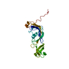
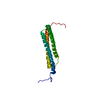
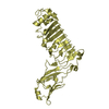
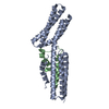
| Title | THE CRYSTAL STRUCTURE OF MUCOSAL ADDRESSIN CELL ADHESION MOLECULE-1 (MADCAM-1) | |||||||||
 Components Components | PROTEIN (MUCOSAL ADDRESSIN CELL ADHESION MOLECULE-1) | |||||||||
 Keywords Keywords | MEMBRANE PROTEIN / CELL ADHESION PROTEIN / MADCAM-1 / IMMUNOGLOBULIN FOLD / I-SET FOLD / CELL ADHESION GLYCOPROTEIN / INTEGRIN RECOGINITION | |||||||||
| Function / homology |  Function and homology information Function and homology informationpositive regulation of lymphocyte migration / integrin binding involved in cell-matrix adhesion / leukocyte tethering or rolling / positive regulation of leukocyte migration / heterotypic cell-cell adhesion / receptor clustering / Integrin cell surface interactions / cell-matrix adhesion / integrin-mediated signaling pathway / Immunoregulatory interactions between a Lymphoid and a non-Lymphoid cell ...positive regulation of lymphocyte migration / integrin binding involved in cell-matrix adhesion / leukocyte tethering or rolling / positive regulation of leukocyte migration / heterotypic cell-cell adhesion / receptor clustering / Integrin cell surface interactions / cell-matrix adhesion / integrin-mediated signaling pathway / Immunoregulatory interactions between a Lymphoid and a non-Lymphoid cell / cell adhesion / immune response / signal transduction / membrane / plasma membrane Similarity search - Function | |||||||||
| Biological species |  Homo sapiens (human) Homo sapiens (human) | |||||||||
| Method |  X-RAY DIFFRACTION / MIR / Resolution: 2.2 Å X-RAY DIFFRACTION / MIR / Resolution: 2.2 Å | |||||||||
 Authors Authors | Tan, K. / Casasnovas, J.M. / Liu, J.H. / Briskin, M.J. / Springer, T.A. / Wang, J.-H. | |||||||||
 Citation Citation | Journal: Structure / Year: 1998 Title: The structure of immunoglobulin superfamily domains 1 and 2 of MAdCAM-1 reveals novel features important for integrin recognition. Authors: Tan, K. / Casasnovas, J.M. / Liu, J.H. / Briskin, M.J. / Springer, T.A. / Wang, J.H. #1: Journal: J.Immunol. / Year: 1996Title: Human Mucosal Addressin Cell Adhesion Molecule-1(Ma Demonstrates Structural and Functional Similarities Alpha4Beta7-Integrin Binding Domains of Murine Madc But Extreme Divergence of Mucin-Like Sequence Authors: Shyjan, A.M. / Bertagnolli, M. / Kenney, C.J. / Briskin, M.J. #2: Journal: Cell(Cambridge,Mass.) / Year: 1994Title: Traffic Signals for Lymphocyte Recirculation and Le Emigration Authors: Springer, T.A. | |||||||||
| History |
|
- Structure visualization
| Structure viewer | Molecule: MolmilJmol/JSmol |
|---|
- Downloads & links
-Download
| PDBx/mmCIF format | 1bqs.cif.gz | 55.1 KB | Display | PDBx/mmCIF format |
|---|---|---|---|---|
| PDB format | pdb1bqs.ent.gz | 39.3 KB | Display | PDB format |
| PDBx/mmJSON format | 1bqs.json.gz | Tree view | PDBx/mmJSON format | |
| Others |  Other downloads Other downloads |
-Validation report
| Arichive directory | https://data.pdbj.org/pub/pdb/validation_reports/bq/1bqsftp://data.pdbj.org/pub/pdb/validation_reports/bq/1bqs | HTTPS FTP |
|---|
-Related structure data
| Similar structure data |
|---|


-Links
 PDBj
PDBj


- Assembly
| Deposited unit | 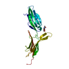
| ||||||||
|---|---|---|---|---|---|---|---|---|---|
| 1 |
| ||||||||
| Unit cell |
|
-Components
| #1: Protein | Mass: 22118.039 Da / Num. of mol.: 1 / Fragment: EXTRACELLULAR REGION WITH TWO IG-LIKE DOMAINS / Source method: isolated from a natural source / Source: (natural) Homo sapiens (human) / Cell: HEMATOPOIETIC AND ENDOTHELIAL CELLS / Plasmid: PBJ5-GS-MADCAM-1 / References: UniProt: Q13477 |
|---|---|
| #2: Sugar | ChemComp-NAG /   Type: D-saccharide, beta linking / Mass: 221.208 Da / Num. of mol.: 1 Type: D-saccharide, beta linking / Mass: 221.208 Da / Num. of mol.: 1Source method: isolated from a genetically manipulated source Formula: C8H15NO6 |
| #3: Water | ChemComp-HOH /  Mass: 18.015 Da / Num. of mol.: 79 / Source method: isolated from a natural source / Formula: H2O Mass: 18.015 Da / Num. of mol.: 79 / Source method: isolated from a natural source / Formula: H2O |
| Compound details | MUCOSAL ADDRESSIN CELL ADHESION MOLECULE-1 (MADCAM-1) IS IMPORTANT IN LYMPHOCYTE HOMING TO MUCOSAL ...MUCOSAL ADDRESSIN CELL ADHESION MOLECULE-1 (MADCAM-1) IS IMPORTANT IN LYMPHOCYTE |
| Has protein modification | Y |
-Experimental details
-Experiment
| Experiment | Method: X-RAY DIFFRACTION / Number of used crystals: 1 |
|---|
- Sample preparation
| Crystal | Density Matthews: 2.63 Å3/Da / Density % sol: 50 % | ||||||||||||||||||
|---|---|---|---|---|---|---|---|---|---|---|---|---|---|---|---|---|---|---|---|
| Crystal grow | Method: vapor diffusion, hanging drop Details: PROTEIN SOLUTION:17 MG/ML. CRYSTALLIZATION SOLUTION: 10% PEG 400, 0.5M LI2SO4, PH 7.5-8.0., VAPOR DIFFUSION, HANGING DROP | ||||||||||||||||||
| Crystal | *PLUS | ||||||||||||||||||
| Crystal grow | *PLUS PH range low: 8 / PH range high: 7.5 | ||||||||||||||||||
| Components of the solutions | *PLUS
|
-Data collection
| Diffraction | Mean temperature: 293 K |
|---|---|
| Diffraction source | Source: ROTATING ANODE / Type: RIGAKU RU200 / Wavelength: 1.5418 |
| Detector | Type: MARRESEARCH / Detector: IMAGE PLATE / Date: Feb 10, 1997 / Details: DOUBLE FOCUSED MIRROR |
| Radiation | Monochromator: NI FILTER / Protocol: SINGLE WAVELENGTH / Monochromatic (M) / Laue (L): M / Scattering type: x-ray |
| Radiation wavelength | Wavelength: 1.5418 Å / Relative weight: 1 |
| Reflection | Resolution: 2.2→15 Å / Num. obs: 11229 / % possible obs: 92.2 % / Observed criterion σ(I): 3 / Redundancy: 7.8 % / Rmerge(I) obs: 0.075 / Net I/σ(I): 22.3 |
| Reflection shell | Resolution: 2.2→2.28 Å / Redundancy: 4.3 % / Rmerge(I) obs: 0.236 / Mean I/σ(I) obs: 6.4 / % possible all: 96.1 |
| Reflection | *PLUS Num. measured all: 87803 |
- Processing
| Software |
| ||||||||||||||||||||||||||||||||||||||||||||||||||||||||||||
|---|---|---|---|---|---|---|---|---|---|---|---|---|---|---|---|---|---|---|---|---|---|---|---|---|---|---|---|---|---|---|---|---|---|---|---|---|---|---|---|---|---|---|---|---|---|---|---|---|---|---|---|---|---|---|---|---|---|---|---|---|---|
| Refinement | Method to determine structure: MIR / Resolution: 2.2→15 Å / σ(F): 2
| ||||||||||||||||||||||||||||||||||||||||||||||||||||||||||||
| Displacement parameters | Biso mean: 44.5 Å2 | ||||||||||||||||||||||||||||||||||||||||||||||||||||||||||||
| Refine analyze | Luzzati coordinate error obs: 0.43 Å | ||||||||||||||||||||||||||||||||||||||||||||||||||||||||||||
| Refinement step | Cycle: LAST / Resolution: 2.2→15 Å
| ||||||||||||||||||||||||||||||||||||||||||||||||||||||||||||
| Refine LS restraints |
| ||||||||||||||||||||||||||||||||||||||||||||||||||||||||||||
| LS refinement shell | Resolution: 2.2→2.3 Å / Total num. of bins used: 8
| ||||||||||||||||||||||||||||||||||||||||||||||||||||||||||||
| Xplor file |
| ||||||||||||||||||||||||||||||||||||||||||||||||||||||||||||
| Software | *PLUS Name: X-PLOR / Version: 3.1 / Classification: refinement | ||||||||||||||||||||||||||||||||||||||||||||||||||||||||||||
| Refinement | *PLUS σ(F): 2 / % reflection Rfree: 10 % / Rfactor Rfree: 0.28 | ||||||||||||||||||||||||||||||||||||||||||||||||||||||||||||
| Solvent computation | *PLUS | ||||||||||||||||||||||||||||||||||||||||||||||||||||||||||||
| Displacement parameters | *PLUS Biso mean: 44.5 Å2 | ||||||||||||||||||||||||||||||||||||||||||||||||||||||||||||
| Refine LS restraints | *PLUS
| ||||||||||||||||||||||||||||||||||||||||||||||||||||||||||||
| LS refinement shell | *PLUS Highest resolution: 2.2 Å / Lowest resolution: 2.3 Å / Rfactor Rfree: 0.369 / % reflection Rfree: 10 % / Rfactor Rwork: 0.327 |
