 Movie
Movie Controller


+ Open data

- Basic information
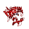
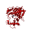
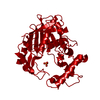
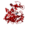
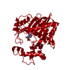
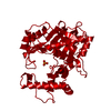
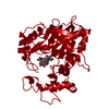
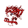
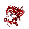
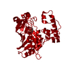
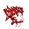
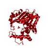
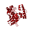
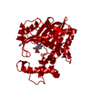
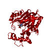
| Entry | Database: PDB / ID: 1bo8 | |||||||||
|---|---|---|---|---|---|---|---|---|---|---|
| Title | THYMIDYLATE SYNTHASE R178T MUTANT | |||||||||
 Components Components | THYMIDYLATE SYNTHASE | |||||||||
 Keywords Keywords | TRANSFERASE / METHYLTRANSFERASE / NUCLEOTIDE BIOSYNTHESIS | |||||||||
| Function / homology |  Function and homology information Function and homology informationthymidylate synthase / thymidylate synthase activity / dTTP biosynthetic process / dTMP biosynthetic process / methylation / cytosol Similarity search - Function | |||||||||
| Biological species |  Lactobacillus casei (bacteria) Lactobacillus casei (bacteria) | |||||||||
| Method |  X-RAY DIFFRACTION / MOLECULAR REPLACEMENT / Resolution: 2.4 Å X-RAY DIFFRACTION / MOLECULAR REPLACEMENT / Resolution: 2.4 Å | |||||||||
 Authors Authors | Morse, R. / Finer-Moore, J. / Stroud, R.M. | |||||||||
 Citation Citation | Journal: Biochemistry / Year: 2000 Title: Energetic contributions of four arginines to phosphate-binding in thymidylate synthase are more than additive and depend on optimization of "effective charge balance". Authors: Morse, R.J. / Kawase, S. / Santi, D.V. / Finer-Moore, J. / Stroud, R.M. #1: Journal: Faseb J. / Year: 1993Title: Stereochemistry of a Multistep/Bipartite Methyl Transfer Reaction: Thymidylate Synthase Authors: Stroud, R.M. / Finer-Moore, J.S. #2: Journal: J.Mol.Biol. / Year: 1993Title: Refined Structures of Substrate-Bound and Phosphate-Bound Thymidylate Synthase from Lactobacillus Casei Authors: Finer-Moore, J.S. / Fauman, E.B. / Foster, P.G. / Perry, K.M. / Santi, D.V. / Stroud, R.M. #3: Journal: Science / Year: 1987Title: Atomic Structure of Thymidylate Synthase: Target for Rational Drug Design Authors: Hardy, L.W. / Finer-Moore, J.S. / Montfort, W.R. / Jones, M.O. / Santi, D.V. / Stroud, R.M. | |||||||||
| History |
|
- Structure visualization
| Structure viewer | Molecule: MolmilJmol/JSmol |
|---|
- Downloads & links
-Download
| PDBx/mmCIF format | 1bo8.cif.gz | 80.1 KB | Display | PDBx/mmCIF format |
|---|---|---|---|---|
| PDB format | pdb1bo8.ent.gz | 59.8 KB | Display | PDB format |
| PDBx/mmJSON format | 1bo8.json.gz | Tree view | PDBx/mmJSON format | |
| Others |  Other downloads Other downloads |
-Validation report
| Arichive directory | https://data.pdbj.org/pub/pdb/validation_reports/bo/1bo8ftp://data.pdbj.org/pub/pdb/validation_reports/bo/1bo8 | HTTPS FTP |
|---|
-Related structure data
| Related structure data |  1bo7C 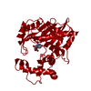 1bp0C 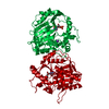 1bp6C 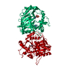 1bpjC  1tdm S: Starting model for refinement C: citing same article ( |
|---|---|
| Similar structure data |


-Links
 PDBj
PDBj

- Assembly
| Deposited unit | 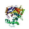
| ||||||||
|---|---|---|---|---|---|---|---|---|---|
| 1 | 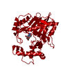
| ||||||||
| Unit cell |
| ||||||||
| Components on special symmetry positions |
|
-Components
| #1: Protein | Mass: 36574.363 Da / Num. of mol.: 1 / Mutation: R178T Source method: isolated from a genetically manipulated source Source: (gene. exp.) Lactobacillus casei (bacteria) / Strain: E. COLI CHI-2913 / Description: SYNTHETIC GENE / Plasmid: PKPTSD / Production host: |
|---|---|
| #2: Chemical | ChemComp-K /   Mass: 39.098 Da / Num. of mol.: 1 / Source method: obtained synthetically / Formula: K Mass: 39.098 Da / Num. of mol.: 1 / Source method: obtained synthetically / Formula: K |
| #3: Chemical | ChemComp-U5P /   Mass: 324.181 Da / Num. of mol.: 1 / Source method: obtained synthetically / Formula: C9H13N2O9P Mass: 324.181 Da / Num. of mol.: 1 / Source method: obtained synthetically / Formula: C9H13N2O9P |
| #4: Water | ChemComp-HOH /  Mass: 18.015 Da / Num. of mol.: 66 / Source method: isolated from a natural source / Formula: H2O Mass: 18.015 Da / Num. of mol.: 66 / Source method: isolated from a natural source / Formula: H2O |
| Has protein modification | N |
| Nonpolymer details | K 1 IS A POSSIBLE POTASSIUM ION THAT LIES ON A MOLECULAR AND CRYSTALLOG |
-Experimental details
-Experiment
| Experiment | Method: X-RAY DIFFRACTION / Number of used crystals: 1 |
|---|
- Sample preparation
| Crystal | Density Matthews: 2.94 Å3/Da / Density % sol: 57 % | ||||||||||||||||||||||||||||||
|---|---|---|---|---|---|---|---|---|---|---|---|---|---|---|---|---|---|---|---|---|---|---|---|---|---|---|---|---|---|---|---|
| Crystal grow | pH: 7 / Details: pH 7.0 | ||||||||||||||||||||||||||||||
| Crystal grow | *PLUS Method: vapor diffusion | ||||||||||||||||||||||||||||||
| Components of the solutions | *PLUS
|
-Data collection
| Diffraction | Mean temperature: 293 K |
|---|---|
| Diffraction source | Source: ROTATING ANODE / Type: RIGAKU RU300 / Wavelength: 1.5418 |
| Detector | Type: SIEMENS / Detector: AREA DETECTOR / Date: Jul 15, 1995 |
| Radiation | Monochromator: GRAPHITE MONOCHROMATOR / Protocol: SINGLE WAVELENGTH / Monochromatic (M) / Laue (L): M / Scattering type: x-ray |
| Radiation wavelength | Wavelength: 1.5418 Å / Relative weight: 1 |
| Reflection | Resolution: 2.4→50 Å / Num. obs: 17212 / % possible obs: 94.3 % / Observed criterion σ(I): 0 / Redundancy: 3.5 % / Biso Wilson estimate: 37.5 Å2 / Rmerge(I) obs: 0.081 / Rsym value: 8.1 / Net I/σ(I): 15.4 |
| Reflection shell | Resolution: 2.4→2.5 Å / Redundancy: 3 % / Mean I/σ(I) obs: 2 / Rsym value: 62 / % possible all: 87.1 |
| Reflection | *PLUS % possible obs: 94.7 % |
| Reflection shell | *PLUS % possible obs: 87.1 % / Rmerge(I) obs: 0.62 |
- Processing
| Software |
| ||||||||||||||||||||||||||||||||||||||||||||||||||||||||||||||||||||||||||||||||
|---|---|---|---|---|---|---|---|---|---|---|---|---|---|---|---|---|---|---|---|---|---|---|---|---|---|---|---|---|---|---|---|---|---|---|---|---|---|---|---|---|---|---|---|---|---|---|---|---|---|---|---|---|---|---|---|---|---|---|---|---|---|---|---|---|---|---|---|---|---|---|---|---|---|---|---|---|---|---|---|---|---|
| Refinement | Method to determine structure: MOLECULAR REPLACEMENT Starting model: 1TDM 1tdm Resolution: 2.4→7 Å / Rfactor Rfree error: 0.007 / Data cutoff high absF: 10000 / Data cutoff low absF: 0.001 / Isotropic thermal model: RESTRAINED / Cross valid method: THROUGHOUT / σ(F): 2 Details: K 1 IS A POSSIBLE POTASSIUM ION THAT LIES ON A MOLECULAR AND CRYSTALLOGRAPHIC TWO-FOLD AXIS.
| ||||||||||||||||||||||||||||||||||||||||||||||||||||||||||||||||||||||||||||||||
| Displacement parameters | Biso mean: 34.4 Å2
| ||||||||||||||||||||||||||||||||||||||||||||||||||||||||||||||||||||||||||||||||
| Refine analyze |
| ||||||||||||||||||||||||||||||||||||||||||||||||||||||||||||||||||||||||||||||||
| Refinement step | Cycle: LAST / Resolution: 2.4→7 Å
| ||||||||||||||||||||||||||||||||||||||||||||||||||||||||||||||||||||||||||||||||
| Refine LS restraints |
| ||||||||||||||||||||||||||||||||||||||||||||||||||||||||||||||||||||||||||||||||
| LS refinement shell | Resolution: 2.4→2.5 Å / Rfactor Rfree error: 0.023 / Total num. of bins used: 8
| ||||||||||||||||||||||||||||||||||||||||||||||||||||||||||||||||||||||||||||||||
| Xplor file |
| ||||||||||||||||||||||||||||||||||||||||||||||||||||||||||||||||||||||||||||||||
| Software | *PLUS Name: X-PLOR / Version: 3.8 / Classification: refinement | ||||||||||||||||||||||||||||||||||||||||||||||||||||||||||||||||||||||||||||||||
| Refinement | *PLUS Rfactor Rfree: 0.247 | ||||||||||||||||||||||||||||||||||||||||||||||||||||||||||||||||||||||||||||||||
| Solvent computation | *PLUS | ||||||||||||||||||||||||||||||||||||||||||||||||||||||||||||||||||||||||||||||||
| Displacement parameters | *PLUS Biso mean: 34 Å2 | ||||||||||||||||||||||||||||||||||||||||||||||||||||||||||||||||||||||||||||||||
| Refine LS restraints | *PLUS
|
