 Movie
Movie Controller


+ Open data

- Basic information
| Entry | Database: PDB / ID: 1bi1 | ||||||
|---|---|---|---|---|---|---|---|
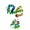
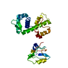
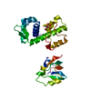
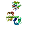
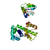
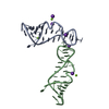
| Title | STRUCTURE OF APO-AND HOLO-DIPHTHERIA TOXIN REPRESSOR | ||||||
 Components Components | DIPHTHERIA TOXIN REPRESSOR | ||||||
 Keywords Keywords | REPRESSOR / TRANSCRIPTION REGULATION / DNA-BINDING / IRON | ||||||
| Function / homology |  Function and homology information Function and homology informationtransition metal ion binding / SH3 domain binding / protein dimerization activity / DNA-binding transcription factor activity / negative regulation of DNA-templated transcription / DNA-templated transcription / DNA binding / identical protein binding / cytoplasm Similarity search - Function | ||||||
| Biological species |  Corynebacterium diphtheriae (bacteria) Corynebacterium diphtheriae (bacteria) | ||||||
| Method |  X-RAY DIFFRACTION / MOLECULAR REPLACEMENT / Resolution: 2.2 Å X-RAY DIFFRACTION / MOLECULAR REPLACEMENT / Resolution: 2.2 Å | ||||||
 Authors Authors | Pohl, E. / Hol, W.G.J. | ||||||
 Citation Citation | Journal: J.Biol.Chem. / Year: 1998 Title: Motion of the DNA-binding domain with respect to the core of the diphtheria toxin repressor (DtxR) revealed in the crystal structures of apo- and holo-DtxR. Authors: Pohl, E. / Holmes, R.K. / Hol, W.G. #1: Journal: Structure / Year: 1995Title: Three-Dimensional Structure of the Diphtheria Toxin Repressor in Complex with Divalent Cation Co-Repressors Authors: Qiu, X. / Verlinde, C.L. / Zhang, S. / Schmitt, M.P. / Holmes, R.K. / Hol, W.G. #2: Journal: Proc.Natl.Acad.Sci.USA / Year: 1992Title: Purification and Characterization of the Diphtheria Toxin Repressor Authors: Schmitt, M.P. / Twiddy, E.M. / Holmes, R.K. | ||||||
| History |
|
- Structure visualization
| Structure viewer | Molecule: MolmilJmol/JSmol |
|---|
- Downloads & links
-Download
| PDBx/mmCIF format | 1bi1.cif.gz | 57.3 KB | Display | PDBx/mmCIF format |
|---|---|---|---|---|
| PDB format | pdb1bi1.ent.gz | 41.2 KB | Display | PDB format |
| PDBx/mmJSON format | 1bi1.json.gz | Tree view | PDBx/mmJSON format | |
| Others |  Other downloads Other downloads |
-Validation report
| Arichive directory | https://data.pdbj.org/pub/pdb/validation_reports/bi/1bi1ftp://data.pdbj.org/pub/pdb/validation_reports/bi/1bi1 | HTTPS FTP |
|---|
-Related structure data


-Links
 PDBj
PDBj- Assembly
| Deposited unit | 
| ||||||||
|---|---|---|---|---|---|---|---|---|---|
| 1 |
| ||||||||
| Unit cell |
|
-Components
| #1: Protein | Mass: 25381.859 Da / Num. of mol.: 1 Source method: isolated from a genetically manipulated source Source: (gene. exp.) Corynebacterium diphtheriae (bacteria) / Strain: C7(-) / Gene: DTXR / Plasmid: PMS298 / Gene (production host): T / Production host: |
|---|---|
| #2: Water | ChemComp-HOH /  Mass: 18.015 Da / Num. of mol.: 171 / Source method: isolated from a natural source / Formula: H2O Mass: 18.015 Da / Num. of mol.: 171 / Source method: isolated from a natural source / Formula: H2O |
| Has protein modification | Y |
-Experimental details
-Experiment
| Experiment | Method: X-RAY DIFFRACTION / Number of used crystals: 1 |
|---|
- Sample preparation
| Crystal | Density Matthews: 2.46 Å3/Da / Density % sol: 57 % | ||||||||||||||||||||||||||||||||||||||||||||||||||||||||
|---|---|---|---|---|---|---|---|---|---|---|---|---|---|---|---|---|---|---|---|---|---|---|---|---|---|---|---|---|---|---|---|---|---|---|---|---|---|---|---|---|---|---|---|---|---|---|---|---|---|---|---|---|---|---|---|---|---|
| Crystal grow | pH: 8.5 / Details: pH 8.5 | ||||||||||||||||||||||||||||||||||||||||||||||||||||||||
| Crystal grow | *PLUS Method: vapor diffusion, hanging drop / pH: 8 | ||||||||||||||||||||||||||||||||||||||||||||||||||||||||
| Components of the solutions | *PLUS
|
-Data collection
| Diffraction | Mean temperature: 100 K |
|---|---|
| Diffraction source | Source: ROTATING ANODE / Type: RIGAKU RUH2R / Wavelength: 1.5418 |
| Detector | Type: RIGAKU RAXIS / Detector: IMAGE PLATE / Date: Aug 1, 1997 / Details: MIRRORS |
| Radiation | Monochromator: MIRRORS / Monochromatic (M) / Laue (L): M / Scattering type: x-ray |
| Radiation wavelength | Wavelength: 1.5418 Å / Relative weight: 1 |
| Reflection | Resolution: 2.2→8 Å / Num. obs: 12830 / % possible obs: 97 % / Observed criterion σ(I): 2 / Redundancy: 7 % / Rmerge(I) obs: 0.05 / Rsym value: 0.05 / Net I/σ(I): 24 |
| Reflection shell | Resolution: 2.2→2.28 Å / Redundancy: 2 % / Rmerge(I) obs: 0.195 / Mean I/σ(I) obs: 4.2 / Rsym value: 0.195 / % possible all: 82 |
| Reflection | *PLUS Num. measured all: 92585 |
| Reflection shell | *PLUS % possible obs: 82 % |
- Processing
| Software |
| ||||||||||||||||||||||||||||||||||||||||||||||||||||||||||||
|---|---|---|---|---|---|---|---|---|---|---|---|---|---|---|---|---|---|---|---|---|---|---|---|---|---|---|---|---|---|---|---|---|---|---|---|---|---|---|---|---|---|---|---|---|---|---|---|---|---|---|---|---|---|---|---|---|---|---|---|---|---|
| Refinement | Method to determine structure: MOLECULAR REPLACEMENT / Resolution: 2.2→8 Å / Cross valid method: THROUGHOUT / σ(F): 2 Details: 2FO-FC ELECTRON DENSITY IS MISSING FOR RESIDUES 1 - 3, 141- 147 AND 199 - 200. THESE RESIDUES ARE ASSUMED TO BE DISORDERED. THE EXPERIMENTAL ELECTRON DENSITY IS WEAK OR MISSING FOR RESIDUES 148 - 226.
| ||||||||||||||||||||||||||||||||||||||||||||||||||||||||||||
| Refinement step | Cycle: LAST / Resolution: 2.2→8 Å
| ||||||||||||||||||||||||||||||||||||||||||||||||||||||||||||
| Refine LS restraints |
| ||||||||||||||||||||||||||||||||||||||||||||||||||||||||||||
| Software | *PLUS Name: X-PLOR / Classification: refinement | ||||||||||||||||||||||||||||||||||||||||||||||||||||||||||||
| Refine LS restraints | *PLUS
|
