 Movie
Movie Controller

[English] 日本語
 Yorodumi
- PDB-1bbp: MOLECULAR STRUCTURE OF THE BILIN BINDING PROTEIN (BBP) FROM PIERI... -
+ Open data

- Basic information
| Entry | Database: PDB / ID: 1bbp | ||||||
|---|---|---|---|---|---|---|---|
| Title | MOLECULAR STRUCTURE OF THE BILIN BINDING PROTEIN (BBP) FROM PIERIS BRASSICAE AFTER REFINEMENT AT 2.0 ANGSTROMS RESOLUTION. | ||||||
 Components Components | BILIN BINDING PROTEIN | ||||||
 Keywords Keywords | BILIN BINDING | ||||||
| Function / homology |  Function and homology information Function and homology informationpigment binding / response to reactive oxygen species / lipid metabolic process / extracellular region / cytoplasm Similarity search - Function | ||||||
| Biological species |  Pieris brassicae (large cabbage white) Pieris brassicae (large cabbage white) | ||||||
| Method |  X-RAY DIFFRACTION / Resolution: 2 Å X-RAY DIFFRACTION / Resolution: 2 Å | ||||||
 Authors Authors | Huber, R. / Schneider, M. / Mayr, I. / Mueller, R. / Deutzmann, R. / Suter, F. / Zuber, H. / Falk, H. / Kayser, H. | ||||||
 Citation Citation | Journal: J.Mol.Biol. / Year: 1987 Title: Molecular structure of the bilin binding protein (BBP) from Pieris brassicae after refinement at 2.0 A resolution. Authors: Huber, R. / Schneider, M. / Mayr, I. / Muller, R. / Deutzmann, R. / Suter, F. / Zuber, H. / Falk, H. / Kayser, H. #1: Journal: J.Mol.Biol. / Year: 1987Title: Crystallization, Crystal Structure Analysis and Preliminary Molecular Model of the Bilin Binding Protein from the Insect Pieris Brassicae Authors: Huber, R. / Schneider, M. / Epp, O. / Mayr, I. / Messerschmidt, A. / Pflugrath, J. / Kayser, H. | ||||||
| History |
|
- Structure visualization
| Structure viewer | Molecule: MolmilJmol/JSmol |
|---|
- Downloads & links
-Download
| PDBx/mmCIF format | 1bbp.cif.gz | 157.7 KB | Display | PDBx/mmCIF format |
|---|---|---|---|---|
| PDB format | pdb1bbp.ent.gz | 127.8 KB | Display | PDB format |
| PDBx/mmJSON format | 1bbp.json.gz | Tree view | PDBx/mmJSON format | |
| Others |  Other downloads Other downloads |
-Validation report
| Arichive directory | https://data.pdbj.org/pub/pdb/validation_reports/bb/1bbpftp://data.pdbj.org/pub/pdb/validation_reports/bb/1bbp | HTTPS FTP |
|---|
-Related structure data
| Similar structure data |
|---|


-Links
 PDBj
PDBj

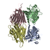
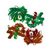
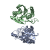
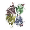
- Assembly
| Deposited unit | 
| ||||||||||||||||||||||||||||
|---|---|---|---|---|---|---|---|---|---|---|---|---|---|---|---|---|---|---|---|---|---|---|---|---|---|---|---|---|---|
| 1 |
| ||||||||||||||||||||||||||||
| Unit cell |
| ||||||||||||||||||||||||||||
| Noncrystallographic symmetry (NCS) | NCS oper:
| ||||||||||||||||||||||||||||
| Details | THE TRANSFORMATIONS PRESENTED ON MTRIX RECORDS BELOW RELATE CHAINS A, B, C, AND D AS FOLLOWS: MTRIX 1: A TO B MTRIX 2: A TO C MTRIX 3: A TO D MTRIX 4: B TO C MTRIX 5: B TO D MTRIX 6: C TO D |
-Components
| #1: Protein | Mass: 19703.029 Da / Num. of mol.: 4 Source method: isolated from a genetically manipulated source Source: (gene. exp.) Pieris brassicae (large cabbage white) / References: UniProt: P09464#2: Chemical | ChemComp-BLV / 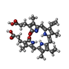  Mass: 582.646 Da / Num. of mol.: 4 / Source method: obtained synthetically / Formula: C33H34N4O6 Mass: 582.646 Da / Num. of mol.: 4 / Source method: obtained synthetically / Formula: C33H34N4O6#3: Water | ChemComp-HOH / |  Mass: 18.015 Da / Num. of mol.: 424 / Source method: isolated from a natural source / Formula: H2O Mass: 18.015 Da / Num. of mol.: 424 / Source method: isolated from a natural source / Formula: H2OHas protein modification | Y | |
|---|
-Experimental details
-Experiment
| Experiment | Method: X-RAY DIFFRACTION |
|---|
- Sample preparation
| Crystal | Density Matthews: 3.26 Å3/Da / Density % sol: 62.25 % | ||||||||||||||||||||||||||||||
|---|---|---|---|---|---|---|---|---|---|---|---|---|---|---|---|---|---|---|---|---|---|---|---|---|---|---|---|---|---|---|---|
| Crystal grow | *PLUS pH: 7 / Method: vapor diffusionDetails: referred to 'Huber, R.', (1987) J.Mol.Biol., 195, 423-434 | ||||||||||||||||||||||||||||||
| Components of the solutions | *PLUS
|
-Data collection
| Radiation | Scattering type: x-ray |
|---|---|
| Radiation wavelength | Relative weight: 1 |
- Processing
| Software | Name: EREF / Classification: refinement | ||||||||||||||||||||||||||||||||||||||||||||||||||||||||||||
|---|---|---|---|---|---|---|---|---|---|---|---|---|---|---|---|---|---|---|---|---|---|---|---|---|---|---|---|---|---|---|---|---|---|---|---|---|---|---|---|---|---|---|---|---|---|---|---|---|---|---|---|---|---|---|---|---|---|---|---|---|---|
| Refinement | Rfactor Rwork: 0.2 / Highest resolution: 2 Å | ||||||||||||||||||||||||||||||||||||||||||||||||||||||||||||
| Refinement step | Cycle: LAST / Highest resolution: 2 Å
| ||||||||||||||||||||||||||||||||||||||||||||||||||||||||||||
| Refine LS restraints |
| ||||||||||||||||||||||||||||||||||||||||||||||||||||||||||||
| Refinement | *PLUS Highest resolution: 2 Å / Num. reflection obs: 51824 / Rfactor Rfree: 0.2 | ||||||||||||||||||||||||||||||||||||||||||||||||||||||||||||
| Solvent computation | *PLUS | ||||||||||||||||||||||||||||||||||||||||||||||||||||||||||||
| Displacement parameters | *PLUS Biso mean: 21 Å2 | ||||||||||||||||||||||||||||||||||||||||||||||||||||||||||||
| Refine LS restraints | *PLUS Type: o_angle_d |
