 Movie
Movie Controller


+ Open data

- Basic information
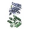
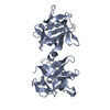
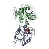
| Entry | Database: PDB / ID: 1ba2 | ||||||
|---|---|---|---|---|---|---|---|
| Title | D67R MUTANT OF D-RIBOSE-BINDING PROTEIN FROM ESCHERICHIA COLI | ||||||
 Components Components | D-RIBOSE-BINDING PROTEIN | ||||||
 Keywords Keywords | TRANSPORT / CHEMOTAXIS / PERIPLASM | ||||||
| Function / homology |  Function and homology information Function and homology informationD-ribose transmembrane transport / monosaccharide binding / positive chemotaxis / ATP-binding cassette (ABC) transporter complex, substrate-binding subunit-containing / transmembrane transport / outer membrane-bounded periplasmic space / membrane Similarity search - Function | ||||||
| Biological species |  | ||||||
| Method |  X-RAY DIFFRACTION / MOLECULAR REPLACEMENT / Resolution: 2.1 Å X-RAY DIFFRACTION / MOLECULAR REPLACEMENT / Resolution: 2.1 Å | ||||||
 Authors Authors | Bjorkman, A.J. / Mowbray, S.L. | ||||||
 Citation Citation | Journal: J.Mol.Biol. / Year: 1998 Title: Multiple open forms of ribose-binding protein trace the path of its conformational change. Authors: Bjorkman, A.J. / Mowbray, S.L. #1: Journal: J.Biol.Chem. / Year: 1994Title: Identical Mutations at Corresponding Positions in Two Homologous Proteins with Nonidentical Effects Authors: Bjorkman, A.J. / Binnie, R.A. / Cole, L.B. / Zhang, H. / Hermodson, M.A. / Mowbray, S.L. #2: Journal: J.Biol.Chem. / Year: 1994Title: Probing Protein-Protein Interactions. The Ribose-Binding Protein in Bacterial Transport and Chemotaxis Authors: Bjorkman, A.J. / Binnie, R.A. / Zhang, H. / Cole, L.B. / Hermodson, M.A. / Mowbray, S.L. #3: Journal: J.Mol.Biol. / Year: 1992Title: 1.7 A X-Ray Structure of the Periplasmic Ribose Receptor from Escherichia Coli Authors: Mowbray, S.L. / Cole, L.B. #4: Journal: Protein Sci. / Year: 1992Title: Functional Mapping of the Surface of Escherichia Coli Ribose-Binding Protein: Mutations that Affect Chemotaxis and Transport Authors: Binnie, R.A. / Zhang, H. / Mowbray, S. / Hermodson, M.A. | ||||||
| History |
|
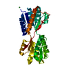
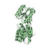
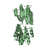
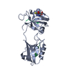
- Structure visualization
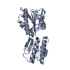
| Structure viewer | Molecule: MolmilJmol/JSmol |
|---|
- Downloads & links
-Download
| PDBx/mmCIF format | 1ba2.cif.gz | 114.4 KB | Display | PDBx/mmCIF format |
|---|---|---|---|---|
| PDB format | pdb1ba2.ent.gz | 89.3 KB | Display | PDB format |
| PDBx/mmJSON format | 1ba2.json.gz | Tree view | PDBx/mmJSON format | |
| Others |  Other downloads Other downloads |
-Validation report
| Arichive directory | https://data.pdbj.org/pub/pdb/validation_reports/ba/1ba2ftp://data.pdbj.org/pub/pdb/validation_reports/ba/1ba2 | HTTPS FTP |
|---|
-Related structure data
| Related structure data |  1urpC  2driS S: Starting model for refinement C: citing same article ( |
|---|---|
| Similar structure data |


-Links
 PDBj
PDBj- Assembly
| Deposited unit | 
| ||||||||
|---|---|---|---|---|---|---|---|---|---|
| 1 | 
| ||||||||
| 2 | 
| ||||||||
| Unit cell |
|
-Components
| #1: Protein | Mass: 28549.529 Da / Num. of mol.: 2 / Mutation: D67R Source method: isolated from a genetically manipulated source Source: (gene. exp.) #2: Water | ChemComp-HOH / |  Mass: 18.015 Da / Num. of mol.: 337 / Source method: isolated from a natural source / Formula: H2O Mass: 18.015 Da / Num. of mol.: 337 / Source method: isolated from a natural source / Formula: H2O |
|---|
-Experimental details
-Experiment
| Experiment | Method: X-RAY DIFFRACTION / Number of used crystals: 1 |
|---|
- Sample preparation
| Crystal | Density Matthews: 2.11 Å3/Da / Density % sol: 41.78 % Description: RESOLUTION LIMITS 8-4 ANGSTROMS IN THE SEARCHES. | ||||||||||||||||||||
|---|---|---|---|---|---|---|---|---|---|---|---|---|---|---|---|---|---|---|---|---|---|
| Crystal grow | Method: vapor diffusion / pH: 7 Details: VAPOR DIFFUSION OF DROPS CONTAINING 7.5 MG/ML PROTEIN AGAINST A RESERVOIR OF 24% PEG4000, 100 MM TRIS-HCL, PH 7., pH 7.0, vapor diffusion | ||||||||||||||||||||
| Crystal grow | *PLUS Method: vapor diffusion / pH: 7 | ||||||||||||||||||||
| Components of the solutions | *PLUS
|
-Data collection
| Diffraction | Mean temperature: 90 K |
|---|---|
| Diffraction source | Source: ROTATING ANODE / Type: RIGAKU RUH3R / Wavelength: 1.5418 |
| Detector | Type: RIGAKU RAXIS IIC / Detector: IMAGE PLATE / Date: Mar 1, 1996 / Details: MIRROR |
| Radiation | Monochromatic (M) / Laue (L): M / Scattering type: x-ray |
| Radiation wavelength | Wavelength: 1.5418 Å / Relative weight: 1 |
| Reflection | Resolution: 2.1→19 Å / Num. obs: 27352 / % possible obs: 94.9 % / Observed criterion σ(I): -3 / Redundancy: 3.36 % / Rmerge(I) obs: 0.038 / Net I/σ(I): 21.3 |
| Reflection shell | Resolution: 2.1→2.2 Å / Redundancy: 2.04 % / Rmerge(I) obs: 0.097 / Mean I/σ(I) obs: 7.6 / % possible all: 70 |
| Reflection shell | *PLUS % possible obs: 70 % |
- Processing
| Software |
| |||||||||||||||||||||
|---|---|---|---|---|---|---|---|---|---|---|---|---|---|---|---|---|---|---|---|---|---|---|
| Refinement | Method to determine structure: MOLECULAR REPLACEMENT Starting model: SEARCH MODELS REPRESENTING ALL NON-HYDROGEN ATOMS FROM DOMAIN 1 (RESIDUES 1-103 AND 236-264) AND DOMAIN 2 (RESIDUES 104-235 AND 265-271) OF PDB ENTRY 2DRI WERE USED SEPARATELY. Resolution: 2.1→19 Å / Cross valid method: THROUGHOUT
| |||||||||||||||||||||
| Refinement step | Cycle: LAST / Resolution: 2.1→19 Å
| |||||||||||||||||||||
| Software | *PLUS Name: REFMAC / Classification: refinement | |||||||||||||||||||||
| Refinement | *PLUS Num. reflection all: 27352 / Rfactor obs: 0.199 | |||||||||||||||||||||
| Solvent computation | *PLUS | |||||||||||||||||||||
| Displacement parameters | *PLUS | |||||||||||||||||||||
| Refine LS restraints | *PLUS
|
