 Movie
Movie Controller


+ Open data

- Basic information
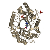
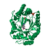
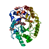
| Entry | Database: PDB / ID: 1b3v | ||||||||||||
|---|---|---|---|---|---|---|---|---|---|---|---|---|---|
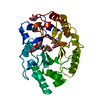
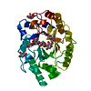
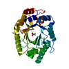
| Title | XYLANASE FROM PENICILLIUM SIMPLICISSIMUM, COMPLEX WITH XYLOSE | ||||||||||||
 Components Components | PROTEIN (XYLANASE) | ||||||||||||
 Keywords Keywords | FAMILY 10 XYLANASE / PENICILLIUM SIMPLICISSIMUM / GLYCOSYL HYDROLASE / SUBSTRATE BINDING | ||||||||||||
| Function / homology |  Function and homology information Function and homology informationendo-1,4-beta-xylanase / endo-1,4-beta-xylanase activity / xylan catabolic process / extracellular region Similarity search - Function | ||||||||||||
| Biological species |  Penicillium simplicissimum (fungus) Penicillium simplicissimum (fungus) | ||||||||||||
| Method |  X-RAY DIFFRACTION / MOLECULAR REPLACEMENT / Resolution: 2.4 Å X-RAY DIFFRACTION / MOLECULAR REPLACEMENT / Resolution: 2.4 Å | ||||||||||||
 Authors Authors | Schmidt, A. / Kratky, C. | ||||||||||||
 Citation Citation | Journal: Biochemistry / Year: 1999 Title: Xylan binding subsite mapping in the xylanase from Penicillium simplicissimum using xylooligosaccharides as cryo-protectant. Authors: Schmidt, A. / Gubitz, G.M. / Kratky, C. #1: Journal: Protein Sci. / Year: 1998Title: Structure of the Xylanase from Penicillium Simplicissimum Authors: Schmidt, A. / Schlacher, A. / Steiner, W. / Schwab, H. / Kratky, C. | ||||||||||||
| History |
|
- Structure visualization
| Structure viewer | Molecule: MolmilJmol/JSmol |
|---|
- Downloads & links
-Download
| PDBx/mmCIF format | 1b3v.cif.gz | 74.4 KB | Display | PDBx/mmCIF format |
|---|---|---|---|---|
| PDB format | pdb1b3v.ent.gz | 55.1 KB | Display | PDB format |
| PDBx/mmJSON format | 1b3v.json.gz | Tree view | PDBx/mmJSON format | |
| Others |  Other downloads Other downloads |
-Validation report
| Arichive directory | https://data.pdbj.org/pub/pdb/validation_reports/b3/1b3vftp://data.pdbj.org/pub/pdb/validation_reports/b3/1b3v | HTTPS FTP |
|---|
-Related structure data
| Related structure data |  1b30C  1b31C  1b3wC  1b3xC  1b3yC  1b3zC 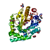 1bg4S S: Starting model for refinement C: citing same article ( |
|---|---|
| Similar structure data |


-Links
 PDBj
PDBj
- Assembly
| Deposited unit | 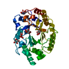
| ||||||||
|---|---|---|---|---|---|---|---|---|---|
| 1 |
| ||||||||
| Unit cell |
|
-Components
| #1: Protein | Mass: 32566.473 Da / Num. of mol.: 1 / Source method: isolated from a natural source / Details: PENICILLIUM SIMPLICISSIMUM (OUDEM.) THOM. / Source: (natural) Penicillium simplicissimum (fungus) / Cellular location: SECRETED / References: UniProt: P56588, endo-1,4-beta-xylanase | ||||||
|---|---|---|---|---|---|---|---|
| #2: Sugar |   Type: D-saccharide, beta linking / Mass: 150.130 Da / Num. of mol.: 3 / Source method: isolated from a natural source / Formula: C5H10O5 Type: D-saccharide, beta linking / Mass: 150.130 Da / Num. of mol.: 3 / Source method: isolated from a natural source / Formula: C5H10O5#3: Sugar | ChemComp-XYS / |   Type: D-saccharide, alpha linking / Mass: 150.130 Da / Num. of mol.: 1 Type: D-saccharide, alpha linking / Mass: 150.130 Da / Num. of mol.: 1Source method: isolated from a genetically manipulated source Formula: C5H10O5 #4: Water | ChemComp-HOH / |  Mass: 18.015 Da / Num. of mol.: 220 / Source method: isolated from a natural source / Formula: H2O Mass: 18.015 Da / Num. of mol.: 220 / Source method: isolated from a natural source / Formula: H2OHas protein modification | Y | |
-Experimental details
-Experiment
| Experiment | Method: X-RAY DIFFRACTION / Number of used crystals: 1 |
|---|
- Sample preparation
| Crystal | Density Matthews: 3.25 Å3/Da / Density % sol: 62 % | ||||||||||||||||||||
|---|---|---|---|---|---|---|---|---|---|---|---|---|---|---|---|---|---|---|---|---|---|
| Crystal grow | pH: 8.4 Details: CRYSTALLIZATION CONDITIONS: PROTEIN WAS CRYSTALLIZED FROM 1.9M (NH4)2SO4, 0.1M TRISHCL PH 8.4 AT 4 C | ||||||||||||||||||||
| Crystal grow | *PLUS Temperature: 4 ℃ / Method: vapor diffusion, hanging drop | ||||||||||||||||||||
| Components of the solutions | *PLUS
|
-Data collection
| Diffraction | Mean temperature: 100 K |
|---|---|
| Diffraction source | Source: ROTATING ANODE / Type: SIEMENS / Wavelength: 1.5418 |
| Detector | Type: MARRESEARCH / Detector: IMAGE PLATE |
| Radiation | Protocol: SINGLE WAVELENGTH / Monochromatic (M) / Laue (L): M / Scattering type: x-ray |
| Radiation wavelength | Wavelength: 1.5418 Å / Relative weight: 1 |
| Reflection | Resolution: 2.4→20 Å / Num. obs: 17527 / % possible obs: 98.9 % / Observed criterion σ(I): -3 / Redundancy: 5 % / Rsym value: 0.185 / Net I/σ(I): 5 |
| Reflection shell | Resolution: 2.4→2.43 Å / Redundancy: 5 % / Mean I/σ(I) obs: 2 / Rsym value: 0.412 / % possible all: 97.9 |
| Reflection | *PLUS Rmerge(I) obs: 0.185 |
| Reflection shell | *PLUS % possible obs: 97.9 % / Rmerge(I) obs: 0.412 |
- Processing
| Software |
| ||||||||||||||||||||
|---|---|---|---|---|---|---|---|---|---|---|---|---|---|---|---|---|---|---|---|---|---|
| Refinement | Method to determine structure: MOLECULAR REPLACEMENT Starting model: PDB ENTRY 1BG4 Resolution: 2.4→20 Å / Data cutoff high absF: 100000 / Data cutoff low absF: 0.001 / Cross valid method: FREE R THROUGHOUT / σ(F): 0 Details: PARAMETER/TOPOLOGY FILES FOR HET GROUPS EXCEPT WATER SELF-SETUP
| ||||||||||||||||||||
| Refinement step | Cycle: LAST / Resolution: 2.4→20 Å
| ||||||||||||||||||||
| LS refinement shell | Resolution: 2.41→2.5 Å / Total num. of bins used: 8
| ||||||||||||||||||||
| Xplor file |
| ||||||||||||||||||||
| Software | *PLUS Name: X-PLOR / Version: 3.851 / Classification: refinement | ||||||||||||||||||||
| Refinement | *PLUS Rfactor obs: 0.211 | ||||||||||||||||||||
| Solvent computation | *PLUS | ||||||||||||||||||||
| Displacement parameters | *PLUS | ||||||||||||||||||||
| LS refinement shell | *PLUS Rfactor obs: 0.273 |
