 Movie
Movie Controller

[English] 日本語
 Yorodumi
- PDB-1b3n: BETA-KETOACYL CARRIER PROTEIN SYNTHASE AS A DRUG TARGET, IMPLICAT... -
+ Open data

- Basic information
| Entry | Database: PDB / ID: 1b3n | ||||||
|---|---|---|---|---|---|---|---|
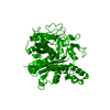
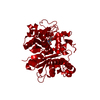
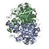
| Title | BETA-KETOACYL CARRIER PROTEIN SYNTHASE AS A DRUG TARGET, IMPLICATIONS FROM THE CRYSTAL STRUCTURE OF A COMPLEX WITH THE INHIBITOR CERULENIN. | ||||||
 Components Components | PROTEIN (KETOACYL ACYL CARRIER PROTEIN SYNTHASE 2) | ||||||
 Keywords Keywords | CONDENSING ENZYMES / FATTY ACID ELONGATION / CERULENIN INHIBITION / LIPID METABOLISM / DRUG DESIGN / DRUG TARGET | ||||||
| Function / homology |  Function and homology information Function and homology informationfatty acid elongation, saturated fatty acid / monounsaturated fatty acid biosynthetic process / beta-ketoacyl-[acyl-carrier-protein] synthase II / 3-oxoacyl-[acyl-carrier-protein] synthase activity / response to cold / fatty acid biosynthetic process / protein homodimerization activity / cytosol Similarity search - Function | ||||||
| Biological species |  | ||||||
| Method |  X-RAY DIFFRACTION / SYNCHROTRON / OTHER / Resolution: 2.65 Å X-RAY DIFFRACTION / SYNCHROTRON / OTHER / Resolution: 2.65 Å | ||||||
 Authors Authors | Moche, M. / Schneider, G. / Edwards, P. / Dehesh, K. / Lindqvist, Y. | ||||||
 Citation Citation | Journal: J.Biol.Chem. / Year: 1999 Title: Structure of the complex between the antibiotic cerulenin and its target, beta-ketoacyl-acyl carrier protein synthase. Authors: Moche, M. / Schneider, G. / Edwards, P. / Dehesh, K. / Lindqvist, Y. | ||||||
| History |
|
- Structure visualization
| Structure viewer | Molecule: MolmilJmol/JSmol |
|---|
- Downloads & links
-Download
| PDBx/mmCIF format | 1b3n.cif.gz | 87.2 KB | Display | PDBx/mmCIF format |
|---|---|---|---|---|
| PDB format | pdb1b3n.ent.gz | 66 KB | Display | PDB format |
| PDBx/mmJSON format | 1b3n.json.gz | Tree view | PDBx/mmJSON format | |
| Others |  Other downloads Other downloads |
-Validation report
| Arichive directory | https://data.pdbj.org/pub/pdb/validation_reports/b3/1b3nftp://data.pdbj.org/pub/pdb/validation_reports/b3/1b3n | HTTPS FTP |
|---|
-Related structure data
| Similar structure data |
|---|


-Links
 PDBj
PDBj

- Assembly
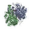
| Deposited unit | 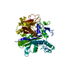
| ||||||||
|---|---|---|---|---|---|---|---|---|---|
| 1 | 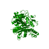
| ||||||||
| Unit cell |
|
-Components
| #1: Protein | Mass: 42958.434 Da / Num. of mol.: 1 Source method: isolated from a genetically manipulated source Source: (gene. exp.) References: UniProt: P0AAI5, beta-ketoacyl-[acyl-carrier-protein] synthase I |
|---|---|
| #2: Chemical | ChemComp-CER / (  Mass: 225.284 Da / Num. of mol.: 1 / Source method: obtained synthetically / Formula: C12H19NO3 / Comment: antifungal, antibiotic*YM Mass: 225.284 Da / Num. of mol.: 1 / Source method: obtained synthetically / Formula: C12H19NO3 / Comment: antifungal, antibiotic*YM |
| #3: Water | ChemComp-HOH /  Mass: 18.015 Da / Num. of mol.: 53 / Source method: isolated from a natural source / Formula: H2O Mass: 18.015 Da / Num. of mol.: 53 / Source method: isolated from a natural source / Formula: H2O |
| Has protein modification | Y |
| Nonpolymer details | THE CERULENIN RESIDUE IS GIVEN CHAIN IDENTIFIER |
-Experimental details
-Experiment
| Experiment | Method: X-RAY DIFFRACTION / Number of used crystals: 2 |
|---|
- Sample preparation
| Crystal | Density Matthews: 2.89 Å3/Da / Density % sol: 57.51 % | ||||||||||||||||||||||||||||||||||||||||||||||||
|---|---|---|---|---|---|---|---|---|---|---|---|---|---|---|---|---|---|---|---|---|---|---|---|---|---|---|---|---|---|---|---|---|---|---|---|---|---|---|---|---|---|---|---|---|---|---|---|---|---|
| Crystal grow | pH: 8 Details: 5.6 MG/ML KAS-CERULENIN COMPLEX IN 0.3M NACL, 25MM TRIS PH8.0, 5MM IMIDAZOLE AND 10% (V/V) GLYCEROL WERE MIXED (2+2UL) WITH A 1000 UL RESERVOIR SOLUTION CONSISTING OF 26% W/V PEG8000 0.1% ...Details: 5.6 MG/ML KAS-CERULENIN COMPLEX IN 0.3M NACL, 25MM TRIS PH8.0, 5MM IMIDAZOLE AND 10% (V/V) GLYCEROL WERE MIXED (2+2UL) WITH A 1000 UL RESERVOIR SOLUTION CONSISTING OF 26% W/V PEG8000 0.1% V/V 2-MERCAPTOETHANOL AND WATER. CRYSTALS APPEARED IN A FEW DAYS | ||||||||||||||||||||||||||||||||||||||||||||||||
| Components of the solutions |
| ||||||||||||||||||||||||||||||||||||||||||||||||
| Crystal grow | *PLUS Temperature: 298 K / Method: vapor diffusion, hanging drop | ||||||||||||||||||||||||||||||||||||||||||||||||
| Components of the solutions | *PLUS
|
-Data collection
| Diffraction | Mean temperature: 298 K |
|---|---|
| Diffraction source | Source: SYNCHROTRON / Site: MAX II  / Beamline: I711 / Wavelength: 0.958 / Beamline: I711 / Wavelength: 0.958 |
| Detector | Type: MARRESEARCH / Detector: IMAGE PLATE / Date: Mar 1, 1998 |
| Radiation | Protocol: SINGLE WAVELENGTH / Monochromatic (M) / Laue (L): M / Scattering type: x-ray |
| Radiation wavelength | Wavelength: 0.958 Å / Relative weight: 1 |
| Reflection | Resolution: 2.65→30 Å / Num. obs: 12600 / % possible obs: 83.4 % / Redundancy: 1.8 % / Rsym value: 0.095 / Net I/σ(I): 5.3 |
| Reflection shell | Resolution: 2.65→2.72 Å / Redundancy: 1.6 % / Mean I/σ(I) obs: 1.6 / Rsym value: 0.325 / % possible all: 71.8 |
| Reflection | *PLUS Num. measured all: 22896 / Rmerge(I) obs: 0.095 |
| Reflection shell | *PLUS % possible obs: 71.8 % / Rmerge(I) obs: 0.325 |
- Processing
| Software |
| |||||||||||||||||||||
|---|---|---|---|---|---|---|---|---|---|---|---|---|---|---|---|---|---|---|---|---|---|---|
| Refinement | Method to determine structure: OTHER / Resolution: 2.65→30 Å / Cross valid method: THROUGHOUT / σ(F): 0 Details: RAMACHANDRAN PLOT, PERCENT OF NON- GLYCINE OR NON-PROLINE RESIDUES IN MOST FAVOURED REGION 88.9% ADDITIONAL ALLOWED REGION 10.5% GENEROUSLY ALLOWED REGION 0.3% DISALLOWED REGION 0.3% BOND ...Details: RAMACHANDRAN PLOT, PERCENT OF NON- GLYCINE OR NON-PROLINE RESIDUES IN MOST FAVOURED REGION 88.9% ADDITIONAL ALLOWED REGION 10.5% GENEROUSLY ALLOWED REGION 0.3% DISALLOWED REGION 0.3% BOND LENGTHS (A) : 0.008 BOND ANGLES ( DEGREES) : 1.7 DIHEDRAL ANGLES (DEGREES) : 26.2 IMPROPER ANGLES (DEGREES) : 1.8
| |||||||||||||||||||||
| Refinement step | Cycle: LAST / Resolution: 2.65→30 Å
| |||||||||||||||||||||
| Software | *PLUS Name: REFMAC / Classification: refinement | |||||||||||||||||||||
| Refinement | *PLUS Num. reflection obs: 11926 / Rfactor obs: 0.213 | |||||||||||||||||||||
| Solvent computation | *PLUS | |||||||||||||||||||||
| Displacement parameters | *PLUS | |||||||||||||||||||||
| Refine LS restraints | *PLUS
|
