 Movie
Movie Controller


+ Open data

- Basic information
| Entry | Database: PDB / ID: 1b0m | |||||||||
|---|---|---|---|---|---|---|---|---|---|---|
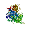
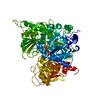
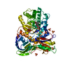
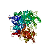
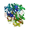
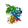
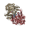
| Title | ACONITASE R644Q:FLUOROCITRATE COMPLEX | |||||||||
 Components Components | PROTEIN (ACONITASE) | |||||||||
 Keywords Keywords | HYDROLASE / ACONITASE R644Q / FLUOROCITRATE COMPLEX | |||||||||
| Function / homology |  Function and homology information Function and homology informationaconitate hydratase / aconitate hydratase activity / tricarboxylic acid cycle / 4 iron, 4 sulfur cluster binding / mitochondrion / metal ion binding / cytosol Similarity search - Function | |||||||||
| Biological species |  | |||||||||
| Method |  X-RAY DIFFRACTION / SYNCHROTRON / MOLECULAR REPLACEMENT / Resolution: 2.5 Å X-RAY DIFFRACTION / SYNCHROTRON / MOLECULAR REPLACEMENT / Resolution: 2.5 Å | |||||||||
 Authors Authors | Lloyd, S.J. / Lauble, H. / Prasad, G.S. / Stout, C.D. | |||||||||
 Citation Citation | Journal: Protein Sci. / Year: 1999 Title: The mechanism of aconitase: 1.8 A resolution crystal structure of the S642a:citrate complex. Authors: Lloyd, S.J. / Lauble, H. / Prasad, G.S. / Stout, C.D. | |||||||||
| History |
|
- Structure visualization
| Structure viewer | Molecule: MolmilJmol/JSmol |
|---|
- Downloads & links
-Download
| PDBx/mmCIF format | 1b0m.cif.gz | 167.8 KB | Display | PDBx/mmCIF format |
|---|---|---|---|---|
| PDB format | pdb1b0m.ent.gz | 129.2 KB | Display | PDB format |
| PDBx/mmJSON format | 1b0m.json.gz | Tree view | PDBx/mmJSON format | |
| Others |  Other downloads Other downloads |
-Validation report
| Arichive directory | https://data.pdbj.org/pub/pdb/validation_reports/b0/1b0mftp://data.pdbj.org/pub/pdb/validation_reports/b0/1b0m | HTTPS FTP |
|---|
-Related structure data
| Related structure data |  1b0jC  1b0kC  1c96C  1c97C  1fghS S: Starting model for refinement C: citing same article ( |
|---|---|
| Similar structure data |


-Links
 PDBj
PDBj


- Assembly
| Deposited unit | 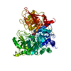
| ||||||||
|---|---|---|---|---|---|---|---|---|---|
| 1 |
| ||||||||
| Unit cell |
|
-Components
| #1: Protein | Mass: 82648.875 Da / Num. of mol.: 1 / Mutation: R644Q Source method: isolated from a genetically manipulated source Details: [4FE-4S] BOUND BY CYS 358, CYS 421, CYS 424 / Source: (gene. exp.)  |
|---|---|
| #2: Chemical | ChemComp-FLC /   Mass: 189.100 Da / Num. of mol.: 1 / Source method: obtained synthetically / Formula: C6H5O7 Mass: 189.100 Da / Num. of mol.: 1 / Source method: obtained synthetically / Formula: C6H5O7 |
| #3: Chemical | ChemComp-SF4 /   Mass: 351.640 Da / Num. of mol.: 1 / Source method: obtained synthetically / Formula: Fe4S4 Mass: 351.640 Da / Num. of mol.: 1 / Source method: obtained synthetically / Formula: Fe4S4 |
| #4: Water | ChemComp-HOH /  Mass: 18.015 Da / Num. of mol.: 341 / Source method: isolated from a natural source / Formula: H2O Mass: 18.015 Da / Num. of mol.: 341 / Source method: isolated from a natural source / Formula: H2O |
| Nonpolymer details | 4 STEREOISOMERS OF FLUOROCITRATE, F ATOM NOT SEEN IN DENSITY, H ATOMS NOT MODELED, CARBOXYL GROUPS ...4 STEREOISOM |
| Sequence details | 1ATQ SWS P16276 1 - 28 NOT IN ATOMS LIST THIS IS RECOMBINANT PORCINE MITOCHONDRIAL ACONITASE - ...1ATQ SWS P16276 1 - 28 NOT IN ATOMS LIST THIS IS RECOMBINAN |
-Experimental details
-Experiment
| Experiment | Method: X-RAY DIFFRACTION / Number of used crystals: 2 |
|---|
- Sample preparation
| Crystal | Density Matthews: 2.81 Å3/Da / Density % sol: 57 % | ||||||||||||||||||||||||||||||||||||||||||||||||
|---|---|---|---|---|---|---|---|---|---|---|---|---|---|---|---|---|---|---|---|---|---|---|---|---|---|---|---|---|---|---|---|---|---|---|---|---|---|---|---|---|---|---|---|---|---|---|---|---|---|
| Crystal grow | pH: 7 / Details: pH 7.0 | ||||||||||||||||||||||||||||||||||||||||||||||||
| Crystal grow | *PLUS Temperature: 26 ℃ / Method: vapor diffusion | ||||||||||||||||||||||||||||||||||||||||||||||||
| Components of the solutions | *PLUS
|
-Data collection
| Diffraction | Mean temperature: 277 K |
|---|---|
| Diffraction source | Source: SYNCHROTRON / Site: SSRL  / Beamline: BL7-1 / Wavelength: 1.08 / Beamline: BL7-1 / Wavelength: 1.08 |
| Detector | Type: MARRESEARCH / Detector: IMAGE PLATE |
| Radiation | Protocol: SINGLE WAVELENGTH / Monochromatic (M) / Laue (L): M / Scattering type: x-ray |
| Radiation wavelength | Wavelength: 1.08 Å / Relative weight: 1 |
| Reflection | Resolution: 2.5→20 Å / Num. obs: 27625 / % possible obs: 84.9 % / Observed criterion σ(I): 0 / Redundancy: 2.3 % / Rsym value: 0.08 / Net I/σ(I): 5.4 |
| Reflection shell | Resolution: 2.5→2.63 Å / Redundancy: 1.9 % / Mean I/σ(I) obs: 1.5 / Rsym value: 0.277 / % possible all: 83 |
- Processing
| Software |
| ||||||||||||||||||||||||||||||||||||||||||||||||||||||||||||
|---|---|---|---|---|---|---|---|---|---|---|---|---|---|---|---|---|---|---|---|---|---|---|---|---|---|---|---|---|---|---|---|---|---|---|---|---|---|---|---|---|---|---|---|---|---|---|---|---|---|---|---|---|---|---|---|---|---|---|---|---|---|
| Refinement | Method to determine structure: MOLECULAR REPLACEMENT Starting model: PDB ENTRY 1FGH Resolution: 2.5→20 Å / Isotropic thermal model: RESTRAINED / σ(F): 0
| ||||||||||||||||||||||||||||||||||||||||||||||||||||||||||||
| Displacement parameters | Biso mean: 21.9 Å2 | ||||||||||||||||||||||||||||||||||||||||||||||||||||||||||||
| Refinement step | Cycle: LAST / Resolution: 2.5→20 Å
| ||||||||||||||||||||||||||||||||||||||||||||||||||||||||||||
| Refine LS restraints |
| ||||||||||||||||||||||||||||||||||||||||||||||||||||||||||||
| Software | *PLUS Name: X-PLOR / Version: 3.8 / Classification: refinement | ||||||||||||||||||||||||||||||||||||||||||||||||||||||||||||
| Refinement | *PLUS Lowest resolution: 20 Å / σ(F): 0 | ||||||||||||||||||||||||||||||||||||||||||||||||||||||||||||
| Solvent computation | *PLUS | ||||||||||||||||||||||||||||||||||||||||||||||||||||||||||||
| Displacement parameters | *PLUS Biso mean: 21.9 Å2 | ||||||||||||||||||||||||||||||||||||||||||||||||||||||||||||
| Refine LS restraints | *PLUS
|
