 Movie
Movie Controller

[English] 日本語
 Yorodumi
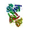
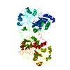
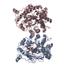
- PDB-1aua: PHOSPHATIDYLINOSITOL TRANSFER PROTEIN SEC14P FROM SACCHAROMYCES C... -
+ Open data

- Basic information
| Entry | Database: PDB / ID: 1aua | ||||||
|---|---|---|---|---|---|---|---|
| Title | PHOSPHATIDYLINOSITOL TRANSFER PROTEIN SEC14P FROM SACCHAROMYCES CEREVISIAE | ||||||
 Components Components | PHOSPHATIDYLINOSITOL TRANSFER PROTEIN SEC14P | ||||||
 Keywords Keywords | PHOSPHOLIPID-BINDING PROTEIN / PERIPHERAL GOLGI MEMBRANE PROTEIN / PHOSPHOLIPID EXCHANGE / GOLGI-DERIVED SECRETORY VESICLE BIOGENESIS | ||||||
| Function / homology |  Function and homology information Function and homology informationnegative regulation of phosphatidylglycerol biosynthetic process / negative regulation of phosphatidylcholine biosynthetic process / phosphatidylcholine intramembrane carrier activity / Golgi vesicle budding / phosphatidylinositol transfer activity / ascospore formation / post-Golgi vesicle-mediated transport / Golgi to vacuole transport / phosphatidylinositol metabolic process / phospholipid transport ...negative regulation of phosphatidylglycerol biosynthetic process / negative regulation of phosphatidylcholine biosynthetic process / phosphatidylcholine intramembrane carrier activity / Golgi vesicle budding / phosphatidylinositol transfer activity / ascospore formation / post-Golgi vesicle-mediated transport / Golgi to vacuole transport / phosphatidylinositol metabolic process / phospholipid transport / Golgi to plasma membrane protein transport / Golgi membrane / Golgi apparatus / cytoplasm / cytosol Similarity search - Function | ||||||
| Biological species |  | ||||||
| Method |  X-RAY DIFFRACTION / SYNCHROTRON / MIR / Resolution: 2.5 Å X-RAY DIFFRACTION / SYNCHROTRON / MIR / Resolution: 2.5 Å | ||||||
 Authors Authors | Sha, B. / Phillips, S.E. / Bankaitis, V.A. / Luo, M. | ||||||
 Citation Citation | Journal: Nature / Year: 1998 Title: Crystal structure of the Saccharomyces cerevisiae phosphatidylinositol-transfer protein. Authors: Sha, B. / Phillips, S.E. / Bankaitis, V.A. / Luo, M. | ||||||
| History |
|
- Structure visualization
| Structure viewer | Molecule: MolmilJmol/JSmol |
|---|
- Downloads & links
-Download
| PDBx/mmCIF format | 1aua.cif.gz | 73.9 KB | Display | PDBx/mmCIF format |
|---|---|---|---|---|
| PDB format | pdb1aua.ent.gz | 54.7 KB | Display | PDB format |
| PDBx/mmJSON format | 1aua.json.gz | Tree view | PDBx/mmJSON format | |
| Others |  Other downloads Other downloads |
-Validation report
| Arichive directory | https://data.pdbj.org/pub/pdb/validation_reports/au/1auaftp://data.pdbj.org/pub/pdb/validation_reports/au/1aua | HTTPS FTP |
|---|
-Related structure data
| Similar structure data |
|---|


-Links
 PDBj
PDBj- Assembly
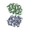
| Deposited unit | 
| ||||||||
|---|---|---|---|---|---|---|---|---|---|
| 1 | 
| ||||||||
| Unit cell |
|
-Components
| #1: Protein | Mass: 34045.578 Da / Num. of mol.: 1 Source method: isolated from a genetically manipulated source Source: (gene. exp.) Gene: SEC14 / Plasmid: PQE-31 / Gene (production host): SEC14 / Production host:  | ||
|---|---|---|---|
| #2: Sugar |   Type: D-saccharide / Mass: 292.369 Da / Num. of mol.: 2 Type: D-saccharide / Mass: 292.369 Da / Num. of mol.: 2Source method: isolated from a genetically manipulated source Formula: C14H28O6 / Comment: detergent*YM #3: Water | ChemComp-HOH / |  Mass: 18.015 Da / Num. of mol.: 53 / Source method: isolated from a natural source / Formula: H2O Mass: 18.015 Da / Num. of mol.: 53 / Source method: isolated from a natural source / Formula: H2O |
-Experimental details
-Experiment
| Experiment | Method: X-RAY DIFFRACTION / Number of used crystals: 1 |
|---|
- Sample preparation
| Crystal | Density Matthews: 3.6 Å3/Da / Density % sol: 70 % | ||||||||||||||||||||||||||||||||||||||||||||||||
|---|---|---|---|---|---|---|---|---|---|---|---|---|---|---|---|---|---|---|---|---|---|---|---|---|---|---|---|---|---|---|---|---|---|---|---|---|---|---|---|---|---|---|---|---|---|---|---|---|---|
| Crystal grow | pH: 7.2 Details: 3% PEG 3350, 1% N-OCTYL B-D-GLUCOPYRANOSIDE, 10 MM HEPES, 150MM KCL, PH 7.2 | ||||||||||||||||||||||||||||||||||||||||||||||||
| Crystal | *PLUS Density % sol: 65 % | ||||||||||||||||||||||||||||||||||||||||||||||||
| Crystal grow | *PLUS Method: vapor diffusion, hanging drop | ||||||||||||||||||||||||||||||||||||||||||||||||
| Components of the solutions | *PLUS
|
-Data collection
| Diffraction | Mean temperature: 100 K |
|---|---|
| Diffraction source | Source: SYNCHROTRON / Site: SSRL  / Beamline: BL7-1 / Wavelength: 1.08 / Beamline: BL7-1 / Wavelength: 1.08 |
| Detector | Type: MARRESEARCH / Detector: IMAGE PLATE / Date: Feb 1, 1997 |
| Radiation | Monochromator: SI(111) / Monochromatic (M) / Laue (L): M / Scattering type: x-ray |
| Radiation wavelength | Wavelength: 1.08 Å / Relative weight: 1 |
| Reflection | Resolution: 2.5→30 Å / Num. obs: 16736 / % possible obs: 90.8 % / Observed criterion σ(I): 2 / Redundancy: 3 % / Biso Wilson estimate: 45 Å2 / Rsym value: 0.051 / Net I/σ(I): 20 |
| Reflection shell | Resolution: 2.5→2.67 Å / Redundancy: 2.1 % / Mean I/σ(I) obs: 8 / Rsym value: 0.265 / % possible all: 80 |
| Reflection | *PLUS Rmerge(I) obs: 0.051 |
| Reflection shell | *PLUS % possible obs: 80 % / Rmerge(I) obs: 0.265 |
- Processing
| Software |
| ||||||||||||||||||||||||||||||||||||||||||||||||||||||||||||
|---|---|---|---|---|---|---|---|---|---|---|---|---|---|---|---|---|---|---|---|---|---|---|---|---|---|---|---|---|---|---|---|---|---|---|---|---|---|---|---|---|---|---|---|---|---|---|---|---|---|---|---|---|---|---|---|---|---|---|---|---|---|
| Refinement | Method to determine structure: MIR / Resolution: 2.5→30 Å / Rfactor Rfree error: 0.01 / Data cutoff high absF: 10000000 / Data cutoff low absF: 0.001 / Cross valid method: THROUGHOUT / σ(F): 2 Details: THE PARAMETER AND TOPOLOGY FILES ARE AVAILABLE FROM DR. B.SHA.
| ||||||||||||||||||||||||||||||||||||||||||||||||||||||||||||
| Displacement parameters | Biso mean: 47.24 Å2 | ||||||||||||||||||||||||||||||||||||||||||||||||||||||||||||
| Refine analyze | Luzzati d res low obs: 30 Å / Luzzati sigma a obs: 0.22 Å | ||||||||||||||||||||||||||||||||||||||||||||||||||||||||||||
| Refinement step | Cycle: LAST / Resolution: 2.5→30 Å
| ||||||||||||||||||||||||||||||||||||||||||||||||||||||||||||
| Refine LS restraints |
| ||||||||||||||||||||||||||||||||||||||||||||||||||||||||||||
| LS refinement shell | Resolution: 2.5→2.61 Å / Rfactor Rfree error: 0.039 / Total num. of bins used: 8
| ||||||||||||||||||||||||||||||||||||||||||||||||||||||||||||
| Xplor file |
| ||||||||||||||||||||||||||||||||||||||||||||||||||||||||||||
| Software | *PLUS Name: X-PLOR / Version: 3.85 / Classification: refinement | ||||||||||||||||||||||||||||||||||||||||||||||||||||||||||||
| Refinement | *PLUS | ||||||||||||||||||||||||||||||||||||||||||||||||||||||||||||
| Solvent computation | *PLUS | ||||||||||||||||||||||||||||||||||||||||||||||||||||||||||||
| Displacement parameters | *PLUS | ||||||||||||||||||||||||||||||||||||||||||||||||||||||||||||
| Refine LS restraints | *PLUS
| ||||||||||||||||||||||||||||||||||||||||||||||||||||||||||||
| LS refinement shell | *PLUS Rfactor obs: 0.354 |
