 Movie
Movie Controller

[English] 日本語
 Yorodumi
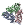
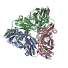
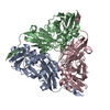
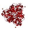
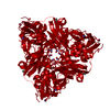
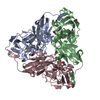
- PDB-1as6: STRUCTURE OF NITRITE BOUND TO OXIDIZED ALCALIGENES FAECALIS NITRI... -
+ Open data

- Basic information
| Entry | Database: PDB / ID: 1as6 | ||||||
|---|---|---|---|---|---|---|---|
| Title | STRUCTURE OF NITRITE BOUND TO OXIDIZED ALCALIGENES FAECALIS NITRITE REDUCTASE AT CRYO TEMPERATURE | ||||||
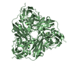
 Components Components | NITRITE REDUCTASE | ||||||
 Keywords Keywords | OXIDOREDUCTASE / NITRITE / COPPER / DENITRIFICATION | ||||||
| Function / homology |  Function and homology information Function and homology informationdenitrification pathway / nitrite reductase (NO-forming) / nitrite reductase (NO-forming) activity / nitrate assimilation / periplasmic space / copper ion binding Similarity search - Function | ||||||
| Biological species |  Alcaligenes faecalis (bacteria) Alcaligenes faecalis (bacteria) | ||||||
| Method |  X-RAY DIFFRACTION / MOLECULAR REPLACEMENT / Resolution: 1.8 Å X-RAY DIFFRACTION / MOLECULAR REPLACEMENT / Resolution: 1.8 Å | ||||||
 Authors Authors | Murphy, M.E.P. / Adman, E.T. / Turley, S. | ||||||
 Citation Citation | Journal: J.Biol.Chem. / Year: 1997 Title: Structure of nitrite bound to copper-containing nitrite reductase from Alcaligenes faecalis. Mechanistic implications. Authors: Murphy, M.E. / Turley, S. / Adman, E.T. #1: Journal: Biochemistry / Year: 1995Title: Structure of Alcaligenes Faecalis Nitrite Reductase and a Copper Site Mutant, M150E, that Contains Zinc Authors: Murphy, M.E. / Turley, S. / Kukimoto, M. / Nishiyama, M. / Horinouchi, S. / Sasaki, H. / Tanokura, M. / Adman, E.T. #2: Journal: Science / Year: 1991Title: The 2.3 Angstrom X-Ray Structure of Nitrite Reductase from Achromobacter Cycloclastes Authors: Godden, J.W. / Turley, S. / Teller, D.C. / Adman, E.T. / Liu, M.Y. / Payne, W.J. / Legall, J. | ||||||
| History |
|
- Structure visualization
| Structure viewer | Molecule: MolmilJmol/JSmol |
|---|
- Downloads & links
-Download
| PDBx/mmCIF format | 1as6.cif.gz | 275.7 KB | Display | PDBx/mmCIF format |
|---|---|---|---|---|
| PDB format | pdb1as6.ent.gz | 223.4 KB | Display | PDB format |
| PDBx/mmJSON format | 1as6.json.gz | Tree view | PDBx/mmJSON format | |
| Others |  Other downloads Other downloads |
-Validation report
| Arichive directory | https://data.pdbj.org/pub/pdb/validation_reports/as/1as6ftp://data.pdbj.org/pub/pdb/validation_reports/as/1as6 | HTTPS FTP |
|---|
-Related structure data
| Related structure data | 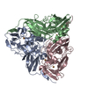 1aq8C 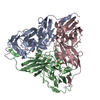 1as7C 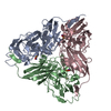 1as8C 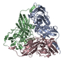 2afnS S: Starting model for refinement C: citing same article ( |
|---|---|
| Similar structure data |

-Links
 PDBj
PDBj

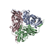
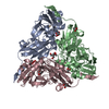
- Assembly
| Deposited unit | 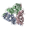
| ||||||||||||
|---|---|---|---|---|---|---|---|---|---|---|---|---|---|
| 1 |
| ||||||||||||
| Unit cell |
| ||||||||||||
| Noncrystallographic symmetry (NCS) | NCS oper:
|
-Components
| #1: Protein | Mass: 37063.887 Da / Num. of mol.: 3 Source method: isolated from a genetically manipulated source Source: (gene. exp.) Alcaligenes faecalis (bacteria) / Strain: S-6 / Cellular location: PERIPLASM / Plasmid: PNIR701 / Cellular location (production host): PERIPLASM / Production host: #2: Chemical | ChemComp-CU /   Mass: 63.546 Da / Num. of mol.: 6 / Source method: obtained synthetically / Formula: Cu Mass: 63.546 Da / Num. of mol.: 6 / Source method: obtained synthetically / Formula: Cu#3: Chemical | 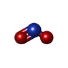  Mass: 46.005 Da / Num. of mol.: 3 / Source method: obtained synthetically / Formula: NO2 Mass: 46.005 Da / Num. of mol.: 3 / Source method: obtained synthetically / Formula: NO2#4: Water | ChemComp-HOH / |  Mass: 18.015 Da / Num. of mol.: 794 / Source method: isolated from a natural source / Formula: H2O Mass: 18.015 Da / Num. of mol.: 794 / Source method: isolated from a natural source / Formula: H2O |
|---|
-Experimental details
-Experiment
| Experiment | Method: X-RAY DIFFRACTION / Number of used crystals: 1 |
|---|
- Sample preparation
| Crystal | Density Matthews: 2.2 Å3/Da / Density % sol: 44 % | ||||||||||||||||||
|---|---|---|---|---|---|---|---|---|---|---|---|---|---|---|---|---|---|---|---|
| Crystal grow | pH: 4.5 / Details: 10% PEG4000, 0.1 SODIUM ACETATE PH 4.5 | ||||||||||||||||||
| Crystal | *PLUS | ||||||||||||||||||
| Crystal grow | *PLUS Method: vapor diffusion, hanging drop / PH range low: 4.8 / PH range high: 4 | ||||||||||||||||||
| Components of the solutions | *PLUS
|
-Data collection
| Diffraction | Mean temperature: 113 K |
|---|---|
| Diffraction source | Source: ROTATING ANODE / Type: RIGAKU RUH2R / Wavelength: 1.5418 |
| Detector | Type: RIGAKU RAXIS IIC / Detector: IMAGE PLATE / Date: May 2, 1995 / Details: MIRRORS |
| Radiation | Monochromator: YALE MIRRORS / Monochromatic (M) / Laue (L): M / Scattering type: x-ray |
| Radiation wavelength | Wavelength: 1.5418 Å / Relative weight: 1 |
| Reflection | Resolution: 1.8→50 Å / Num. obs: 70076 / % possible obs: 81 % / Observed criterion σ(I): 0 / Redundancy: 2.9 % / Rmerge(I) obs: 0.054 / Net I/σ(I): 13.6 |
| Reflection shell | Resolution: 1.8→2 Å / Redundancy: 1.5 % / Rmerge(I) obs: 0.108 / Mean I/σ(I) obs: 3.9 / % possible all: 0.44 |
| Reflection shell | *PLUS % possible obs: 44 % / Num. unique obs: 10128 |
- Processing
| Software |
| ||||||||||||||||||||||||||||||||||||||||||||||||||||||||||||||||||||||||||||||||
|---|---|---|---|---|---|---|---|---|---|---|---|---|---|---|---|---|---|---|---|---|---|---|---|---|---|---|---|---|---|---|---|---|---|---|---|---|---|---|---|---|---|---|---|---|---|---|---|---|---|---|---|---|---|---|---|---|---|---|---|---|---|---|---|---|---|---|---|---|---|---|---|---|---|---|---|---|---|---|---|---|---|
| Refinement | Method to determine structure: MOLECULAR REPLACEMENT Starting model: PDB ENTRY 2AFN Resolution: 1.8→10 Å / Data cutoff high absF: 100000 / Data cutoff low absF: 0.1 / Isotropic thermal model: RESTRAINED / σ(F): 0 /
| ||||||||||||||||||||||||||||||||||||||||||||||||||||||||||||||||||||||||||||||||
| Displacement parameters | Biso mean: 19.12 Å2 | ||||||||||||||||||||||||||||||||||||||||||||||||||||||||||||||||||||||||||||||||
| Refinement step | Cycle: LAST / Resolution: 1.8→10 Å
| ||||||||||||||||||||||||||||||||||||||||||||||||||||||||||||||||||||||||||||||||
| Refine LS restraints |
| ||||||||||||||||||||||||||||||||||||||||||||||||||||||||||||||||||||||||||||||||
| LS refinement shell | Resolution: 1.8→1.98 Å / Total num. of bins used: 8 /
| ||||||||||||||||||||||||||||||||||||||||||||||||||||||||||||||||||||||||||||||||
| Software | *PLUS Name: X-PLOR / Version: 3.1 / Classification: refinement | ||||||||||||||||||||||||||||||||||||||||||||||||||||||||||||||||||||||||||||||||
| Refine LS restraints | *PLUS
|
