 Movie
Movie Controller

[English] 日本語
 Yorodumi
- PDB-1aqe: CRYSTAL STRUCTURE OF THE Y73E MUTANT OF CYTOCHROME C OF CLASS III... -
+ Open data

- Basic information
| Entry | Database: PDB / ID: 1aqe | ||||||
|---|---|---|---|---|---|---|---|
| Title | CRYSTAL STRUCTURE OF THE Y73E MUTANT OF CYTOCHROME C OF CLASS III (AMBLER) 26 KD | ||||||
 Components Components | CYTOCHROME C3 | ||||||
 Keywords Keywords | ELECTRON TRANSPORT / OCTAHEME CYTOCHROME / POINT MUTANT | ||||||
| Function / homology |  Function and homology information Function and homology informationanaerobic respiration / periplasmic space / electron transfer activity / heme binding / metal ion binding Similarity search - Function | ||||||
| Biological species |  Desulfomicrobium norvegicum (bacteria) Desulfomicrobium norvegicum (bacteria) | ||||||
| Method |  X-RAY DIFFRACTION / MOLECULAR REPLACEMENT / Resolution: 2.2 Å X-RAY DIFFRACTION / MOLECULAR REPLACEMENT / Resolution: 2.2 Å | ||||||
 Authors Authors | Czjzek, M. / Haser, R. | ||||||
 Citation Citation | Journal: Biochemistry / Year: 1998 Title: Structural and kinetic studies of the Y73E mutant of octaheme cytochrome c3 (Mr = 26 000) from Desulfovibrio desulfuricans Norway. Authors: Aubert, C. / Giudici-Orticoni, M.T. / Czjzek, M. / Haser, R. / Bruschi, M. / Dolla, A. #1: Journal: J.Biol.Chem. / Year: 1997Title: A Single Mutation in the Heme 4 Environment of Desulfovibrio Desulfuricans Norway Cytochrome C3 (Mr 26,000) Greatly Affects the Molecule Reactivity Authors: Aubert, C. / Leroy, G. / Bruschi, M. / Wall, J.D. / Dolla, A. #2: Journal: Structure / Year: 1996Title: Crystal Structure of a Dimeric Octaheme Cytochrome C3 (Mr 26,000) from Desulfovibrio Desulfuricans Norway Authors: Czjzek, M. / Guerlesquin, F. / Bruschi, M. / Haser, R. | ||||||
| History |
|
- Structure visualization
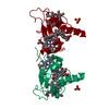
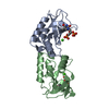
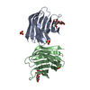
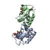
| Structure viewer | Molecule: MolmilJmol/JSmol |
|---|
- Downloads & links
-Download
| PDBx/mmCIF format | 1aqe.cif.gz | 51.1 KB | Display | PDBx/mmCIF format |
|---|---|---|---|---|
| PDB format | pdb1aqe.ent.gz | 37.1 KB | Display | PDB format |
| PDBx/mmJSON format | 1aqe.json.gz | Tree view | PDBx/mmJSON format | |
| Others |  Other downloads Other downloads |
-Validation report
| Summary document | 1aqe_validation.pdf.gz | 1.6 MB | Display | wwPDB validaton report |
|---|---|---|---|---|
| Full document | 1aqe_full_validation.pdf.gz | 1.6 MB | Display | |
| Data in XML | 1aqe_validation.xml.gz | 8.2 KB | Display | |
| Data in CIF | 1aqe_validation.cif.gz | 10.7 KB | Display | |
| Arichive directory | https://data.pdbj.org/pub/pdb/validation_reports/aq/1aqeftp://data.pdbj.org/pub/pdb/validation_reports/aq/1aqe | HTTPS FTP |
-Related structure data
| Related structure data |  1czjS S: Starting model for refinement |
|---|---|
| Similar structure data |


-Links
 PDBj
PDBj
- Assembly
| Deposited unit | 
| ||||||||
|---|---|---|---|---|---|---|---|---|---|
| 1 | 
| ||||||||
| Unit cell |
|
-Components
| #1: Protein | Mass: 12506.900 Da / Num. of mol.: 1 / Mutation: Y73E Source method: isolated from a genetically manipulated source Details: MR 26000 / Source: (gene. exp.) Desulfomicrobium norvegicum (bacteria) / Strain: Norway 4Description: SULFATE REDUCING BACTERIA, ALSO KNOWN AS DESULFOMICROBIUM BACULATUM Plasmid: PJRD215 / Production host: Desulfovibrio desulfuricans (bacteria) / References: UniProt: P38554 | ||||
|---|---|---|---|---|---|
| #2: Chemical | ChemComp-SO4 /   Mass: 96.063 Da / Num. of mol.: 1 / Source method: obtained synthetically / Formula: SO4 Mass: 96.063 Da / Num. of mol.: 1 / Source method: obtained synthetically / Formula: SO4 | ||||
| #3: Chemical | ChemComp-HEM /   Mass: 616.487 Da / Num. of mol.: 4 / Source method: obtained synthetically / Formula: C34H32FeN4O4 Mass: 616.487 Da / Num. of mol.: 4 / Source method: obtained synthetically / Formula: C34H32FeN4O4#4: Water | ChemComp-HOH / |  Mass: 18.015 Da / Num. of mol.: 102 / Source method: isolated from a natural source / Formula: H2O Mass: 18.015 Da / Num. of mol.: 102 / Source method: isolated from a natural source / Formula: H2OHas protein modification | Y | |
-Experimental details
-Experiment
| Experiment | Method: X-RAY DIFFRACTION / Number of used crystals: 1 |
|---|
- Sample preparation
| Crystal | Density Matthews: 3.53 Å3/Da / Density % sol: 65 % | |||||||||||||||||||||||||
|---|---|---|---|---|---|---|---|---|---|---|---|---|---|---|---|---|---|---|---|---|---|---|---|---|---|---|
| Crystal grow | pH: 7.6 Details: THE PROTEIN WAS CRYSTALLIZED FROM 2.4M AMMONIUM SULFATE, 100MM TRIS PH 7.6 | |||||||||||||||||||||||||
| Crystal | *PLUS | |||||||||||||||||||||||||
| Crystal grow | *PLUS Temperature: 4 ℃ / Method: vapor diffusion, hanging drop / Details: Czjzek, M., (1992) J. Mol. Biol., 228, 995. | |||||||||||||||||||||||||
| Components of the solutions | *PLUS
|
-Data collection
| Diffraction | Mean temperature: 293 K |
|---|---|
| Diffraction source | Source: ROTATING ANODE / Type: RIGAKU / Wavelength: 1.5418 |
| Detector | Type: MARRESEARCH / Detector: IMAGE PLATE / Date: Mar 20, 1997 |
| Radiation | Monochromatic (M) / Laue (L): M / Scattering type: x-ray |
| Radiation wavelength | Wavelength: 1.5418 Å / Relative weight: 1 |
| Reflection | Resolution: 2.2→25 Å / Num. obs: 9880 / % possible obs: 95.7 % / Observed criterion σ(I): 1 / Redundancy: 5.2 % / Biso Wilson estimate: 20.97 Å2 / Rmerge(I) obs: 0.096 |
| Reflection shell | Resolution: 2.2→2.25 Å / Redundancy: 4 % / Rmerge(I) obs: 0.35 / % possible all: 91.1 |
| Reflection | *PLUS Num. measured all: 53789 |
- Processing
| Software |
| ||||||||||||||||||||||||||||||||||||||||||||||||||||||||||||
|---|---|---|---|---|---|---|---|---|---|---|---|---|---|---|---|---|---|---|---|---|---|---|---|---|---|---|---|---|---|---|---|---|---|---|---|---|---|---|---|---|---|---|---|---|---|---|---|---|---|---|---|---|---|---|---|---|---|---|---|---|---|
| Refinement | Method to determine structure: MOLECULAR REPLACEMENT Starting model: PDB ENTRY 1CZJ Resolution: 2.2→22 Å / Data cutoff high absF: 10000000 / Data cutoff low absF: 0.001 / σ(F): 1
| ||||||||||||||||||||||||||||||||||||||||||||||||||||||||||||
| Displacement parameters | Biso mean: 21.32 Å2 | ||||||||||||||||||||||||||||||||||||||||||||||||||||||||||||
| Refine analyze | Luzzati coordinate error obs: 0.2 Å | ||||||||||||||||||||||||||||||||||||||||||||||||||||||||||||
| Refinement step | Cycle: LAST / Resolution: 2.2→22 Å
| ||||||||||||||||||||||||||||||||||||||||||||||||||||||||||||
| Refine LS restraints |
| ||||||||||||||||||||||||||||||||||||||||||||||||||||||||||||
| LS refinement shell | Resolution: 2.2→2.3 Å / Total num. of bins used: 8
| ||||||||||||||||||||||||||||||||||||||||||||||||||||||||||||
| Xplor file |
| ||||||||||||||||||||||||||||||||||||||||||||||||||||||||||||
| Software | *PLUS Name: X-PLOR / Version: 3.843 / Classification: refinement | ||||||||||||||||||||||||||||||||||||||||||||||||||||||||||||
| Refine LS restraints | *PLUS
|
