 Movie
Movie Controller

[English] 日本語
 Yorodumi
- PDB-1ahs: CRYSTAL STRUCTURE OF THE TOP DOMAIN OF AFRICAN HORSE SICKNESS VIR... -
+ Open data

- Basic information
| Entry | Database: PDB / ID: 1ahs | ||||||
|---|---|---|---|---|---|---|---|
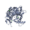
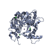
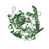
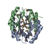
| Title | CRYSTAL STRUCTURE OF THE TOP DOMAIN OF AFRICAN HORSE SICKNESS VIRUS VP7 | ||||||
 Components Components | AFRICAN HORSE SICKNESS VIRUS (SEROTYPE 4) VP7 | ||||||
 Keywords Keywords | VIRAL PROTEIN / CORE PROTEIN / GLYCOPROTEIN / COAT PROTEIN (VIRAL) | ||||||
| Function / homology |  Function and homology information Function and homology informationviral outer capsid / host cell surface receptor binding / fusion of virus membrane with host plasma membrane / viral envelope / structural molecule activity Similarity search - Function | ||||||
| Biological species |  African horsesickness virus African horsesickness virus | ||||||
| Method |  X-RAY DIFFRACTION / SYNCHROTRON / MIR/molecular replacement / Resolution: 2.3 Å X-RAY DIFFRACTION / SYNCHROTRON / MIR/molecular replacement / Resolution: 2.3 Å | ||||||
 Authors Authors | Stuart, D. / Gouet, P. | ||||||
 Citation Citation | Journal: J.Virol. / Year: 1996 Title: Crystal structure of the top domain of African horse sickness virus VP7: comparisons with bluetongue virus VP7. Authors: Basak, A.K. / Gouet, P. / Grimes, J. / Roy, P. / Stuart, D. #1: Journal: Nature / Year: 1995Title: The Crystal Structure of Bluetongue Virus Vp7 Authors: Grimes, J. / Basak, A.K. / Roy, P. / Stuart, D. | ||||||
| History |
|
- Structure visualization
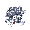
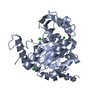
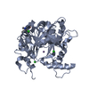
| Structure viewer | Molecule: MolmilJmol/JSmol |
|---|
- Downloads & links
-Download
| PDBx/mmCIF format | 1ahs.cif.gz | 79.9 KB | Display | PDBx/mmCIF format |
|---|---|---|---|---|
| PDB format | pdb1ahs.ent.gz | 62.4 KB | Display | PDB format |
| PDBx/mmJSON format | 1ahs.json.gz | Tree view | PDBx/mmJSON format | |
| Others |  Other downloads Other downloads |
-Validation report
| Arichive directory | https://data.pdbj.org/pub/pdb/validation_reports/ah/1ahsftp://data.pdbj.org/pub/pdb/validation_reports/ah/1ahs | HTTPS FTP |
|---|
-Related structure data
| Related structure data |  1bvpS S: Starting model for refinement |
|---|---|
| Similar structure data |

-Links
 PDBj
PDBj


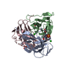
- Assembly
| Deposited unit | 
| ||||||||||||
|---|---|---|---|---|---|---|---|---|---|---|---|---|---|
| 1 |
| ||||||||||||
| Unit cell |
| ||||||||||||
| Noncrystallographic symmetry (NCS) | NCS oper:
|
-Components
| #1: Protein | Mass: 13443.129 Da / Num. of mol.: 3 / Fragment: TOP DOMAIN FRAGMENT / Source method: isolated from a natural source / Source: (natural) African horsesickness virus / Genus: Orbivirus / References: UniProt: P36325#2: Water | ChemComp-HOH / |  Mass: 18.015 Da / Num. of mol.: 61 / Source method: isolated from a natural source / Formula: H2O Mass: 18.015 Da / Num. of mol.: 61 / Source method: isolated from a natural source / Formula: H2OCompound details | THE TRIMERIC FRAGMENT OF AFRICAN HORSESICKNESS VIRUS CAN BE SUPERIMPOSED WITH THE 'TOP DOMAIN' OF ...THE TRIMERIC FRAGMENT OF AFRICAN HORSESICKN | |
|---|
-Experimental details
-Experiment
| Experiment | Method: X-RAY DIFFRACTION / Number of used crystals: 1 |
|---|
- Sample preparation
| Crystal | Density Matthews: 4.42 Å3/Da / Density % sol: 64 % | ||||||||||||||||||||||||||||||
|---|---|---|---|---|---|---|---|---|---|---|---|---|---|---|---|---|---|---|---|---|---|---|---|---|---|---|---|---|---|---|---|
| Crystal grow | *PLUS Temperature: 20 ℃ / pH: 7.5 / Method: vapor diffusion, sitting drop | ||||||||||||||||||||||||||||||
| Components of the solutions | *PLUS
|
-Data collection
| Diffraction |
| |||||||||||||||
|---|---|---|---|---|---|---|---|---|---|---|---|---|---|---|---|---|
| Diffraction source |
| |||||||||||||||
| Detector |
| |||||||||||||||
| Radiation |
| |||||||||||||||
| Radiation wavelength |
| |||||||||||||||
| Reflection | Resolution: 2.3→30 Å / Num. obs: 29456 / % possible obs: 91 % / Observed criterion σ(I): -2 / Redundancy: 3.8 % / Rmerge(I) obs: 0.089 |

- Processing
| Software |
| ||||||||||||||||||||||||||||||||||||||||||||||||||||||||||||
|---|---|---|---|---|---|---|---|---|---|---|---|---|---|---|---|---|---|---|---|---|---|---|---|---|---|---|---|---|---|---|---|---|---|---|---|---|---|---|---|---|---|---|---|---|---|---|---|---|---|---|---|---|---|---|---|---|---|---|---|---|---|
| Refinement | Method to determine structure: MIR/molecular replacement Starting model: 1BVP Resolution: 2.3→15 Å / σ(F): 0 Details: THE REFINEMENT WAS CARRIED OUT AGAINST A MERGE OF THE IN-HOUSE AND SYNCHROTRON DATA. THE THREE SUBUNITS WERE REFINED INDEPENDENTLY. THE WATER MOLECULE HOH 1 LOCATED ALONG THE PSEUDO ...Details: THE REFINEMENT WAS CARRIED OUT AGAINST A MERGE OF THE IN-HOUSE AND SYNCHROTRON DATA. THE THREE SUBUNITS WERE REFINED INDEPENDENTLY. THE WATER MOLECULE HOH 1 LOCATED ALONG THE PSEUDO MOLECULAR THREE-FOLD AXIS HAS AN UNLIKELY LOW TEMPERATURE FACTOR OF 2.00 A**2 AND MAY BE A CHLORIDE ION. THE WATER MOLECULE HOH 1 LOCATED ALONG THE PSEUDO MOLECULAR THREE-FOLD AXIS HAS AN UNLIKELY LOW TEMPERATURE FACTOR OF 2.00 A**2 AND MAY BE A CHLORIDE ION.
| ||||||||||||||||||||||||||||||||||||||||||||||||||||||||||||
| Displacement parameters | Biso mean: 35 Å2 | ||||||||||||||||||||||||||||||||||||||||||||||||||||||||||||
| Refinement step | Cycle: LAST / Resolution: 2.3→15 Å
| ||||||||||||||||||||||||||||||||||||||||||||||||||||||||||||
| Refine LS restraints |
| ||||||||||||||||||||||||||||||||||||||||||||||||||||||||||||
| Software | *PLUS Name: X-PLOR / Classification: refinement | ||||||||||||||||||||||||||||||||||||||||||||||||||||||||||||
| Refinement | *PLUS | ||||||||||||||||||||||||||||||||||||||||||||||||||||||||||||
| Solvent computation | *PLUS | ||||||||||||||||||||||||||||||||||||||||||||||||||||||||||||
| Displacement parameters | *PLUS |
