 Movie
Movie Controller

[English] 日本語
 Yorodumi
- PDB-1afb: STRUCTURAL BASIS OF GALACTOSE RECOGNITION IN C-TYPE ANIMAL LECTINS -
+ Open data

- Basic information
| Entry | Database: PDB / ID: 1afb | ||||||
|---|---|---|---|---|---|---|---|
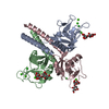
| Title | STRUCTURAL BASIS OF GALACTOSE RECOGNITION IN C-TYPE ANIMAL LECTINS | ||||||
 Components Components | MANNOSE-BINDING PROTEIN-A | ||||||
 Keywords Keywords | LECTIN / C-TYPE LECTIN / CALCIUM-BINDING PROTEIN | ||||||
| Function / homology |  Function and homology information Function and homology informationcalcium-dependent carbohydrate binding / complement activation, lectin pathway / oligosaccharide binding / : / surfactant homeostasis / phosphatidylinositol-4-phosphate binding / protein homotrimerization / D-mannose binding / polysaccharide binding / complement activation, classical pathway ...calcium-dependent carbohydrate binding / complement activation, lectin pathway / oligosaccharide binding / : / surfactant homeostasis / phosphatidylinositol-4-phosphate binding / protein homotrimerization / D-mannose binding / polysaccharide binding / complement activation, classical pathway / multivesicular body / positive regulation of phagocytosis / calcium-dependent protein binding / protease binding / defense response to Gram-positive bacterium / calcium ion binding / protein homodimerization activity / : / identical protein binding Similarity search - Function | ||||||
| Biological species |  | ||||||
| Method |  X-RAY DIFFRACTION / Resolution: 1.9 Å X-RAY DIFFRACTION / Resolution: 1.9 Å | ||||||
 Authors Authors | Kolatkar, A.R. / Weis, W.I. | ||||||
 Citation Citation | Journal: J.Biol.Chem. / Year: 1996 Title: Structural basis of galactose recognition by C-type animal lectins. Authors: Kolatkar, A.R. / Weis, W.I. #1: Journal: Structure / Year: 1994Title: Trimeric Structure of a C-Type Mannose-Binding Protein Authors: Weis, W.I. / Drickamer, K. #2: Journal: J.Biol.Chem. / Year: 1994Title: Binding of Sugar Ligands to Ca+2-Dependent Animal Lectins II. Generation of High-Affinity Galactose Binding by Site-Directed Mutagenesis Authors: Iobst, S.T. / Drickamer, K. | ||||||
| History |
|
- Structure visualization
| Structure viewer | Molecule: MolmilJmol/JSmol |
|---|
- Downloads & links
-Download
| PDBx/mmCIF format | 1afb.cif.gz | 114 KB | Display | PDBx/mmCIF format |
|---|---|---|---|---|
| PDB format | pdb1afb.ent.gz | 87 KB | Display | PDB format |
| PDBx/mmJSON format | 1afb.json.gz | Tree view | PDBx/mmJSON format | |
| Others |  Other downloads Other downloads |
-Validation report
| Arichive directory | https://data.pdbj.org/pub/pdb/validation_reports/af/1afbftp://data.pdbj.org/pub/pdb/validation_reports/af/1afb | HTTPS FTP |
|---|
-Related structure data


-Links
 PDBj
PDBj


- Assembly
| Deposited unit | 
| ||||||||||||
|---|---|---|---|---|---|---|---|---|---|---|---|---|---|
| 1 |
| ||||||||||||
| Unit cell |
| ||||||||||||
| Atom site foot note | 1: CIS PROLINE - PRO 1 186 / 2: CIS PROLINE - PRO 2 186 / 3: CIS PROLINE - PRO 3 186 | ||||||||||||
| Noncrystallographic symmetry (NCS) | NCS oper:
| ||||||||||||
| Details | MTRIX THE TRANSFORMATIONS PRESENTED ON MTRIX RECORDS BELOW DESCRIBE NON-CRYSTALLOGRAPHIC RELATIONSHIPS AMONG THE VARIOUS DOMAINS IN THIS ENTRY. APPLYING THE APPROPRIATE MTRIX TRANSFORMATION TO THE RESIDUES LISTED FIRST WILL YIELD APPROXIMATE COORDINATES FOR THE RESIDUES LISTED SECOND. APPLIED TO TRANSFORMED TO MTRIX RESIDUES RESIDUES RMSD M1 2 73 .. 2 226 1 73 .. 1 226 0.679 M2 3 73 .. 3 226 1 73 .. 1 226 0.329 THE TRANSFORMATIONS WERE DERIVED BY LEAST-SQUARES SUPERPOSITION OF THE MAIN CHAIN N, CA, C, O, CB, WHERE PRESENT, OF RESIDUES 73 TO 226. |
-Components
| #1: Protein | Mass: 17025.295 Da / Num. of mol.: 3 / Fragment: CLOSTRIPAIN FRAGMENT (RESIDUES 73 - 226) Mutation: E185Q, N187D, H189W, G190Y, S191G, INS(H192, G193, L194, G195, G196) Source method: isolated from a genetically manipulated source Details: PH 8.0, DATA COLLECTED AT 100K, 20% 2-METHYL,2-4 PENTANEDIOL (CRYOPROTECTANT) Source: (gene. exp.) Description: THE BACTERIALLY EXPRESSED MATERIAL IS DIGESTED WITH CLOSTRIPAIN TO PRODUCE THE PROTEIN USED IN THE CRYSTAL STRUCTURE ANALYSIS Plasmid: PINIIIOMPA2 / Production host:  #2: Sugar |   Type: D-saccharide, beta linking / Mass: 221.208 Da / Num. of mol.: 3 Type: D-saccharide, beta linking / Mass: 221.208 Da / Num. of mol.: 3Source method: isolated from a genetically manipulated source Formula: C8H15NO6 #3: Chemical | ChemComp-CA /   Mass: 40.078 Da / Num. of mol.: 9 / Source method: obtained synthetically / Formula: Ca Mass: 40.078 Da / Num. of mol.: 9 / Source method: obtained synthetically / Formula: Ca#4: Chemical |   Mass: 35.453 Da / Num. of mol.: 2 / Source method: obtained synthetically / Formula: Cl Mass: 35.453 Da / Num. of mol.: 2 / Source method: obtained synthetically / Formula: Cl#5: Water | ChemComp-HOH / |  Mass: 18.015 Da / Num. of mol.: 371 / Source method: isolated from a natural source / Formula: H2O Mass: 18.015 Da / Num. of mol.: 371 / Source method: isolated from a natural source / Formula: H2OHas protein modification | Y | Sequence details | THERE IS AN INSERTION AFTER RESIDUE SER 191. RESIDUES ARE NUMBERED CONSECUTIV | |
|---|
-Experimental details
-Experiment
| Experiment | Method: X-RAY DIFFRACTION |
|---|
- Sample preparation
| Crystal | Density Matthews: 3.14 Å3/Da / Density % sol: 60.87 % | ||||||||||||||||||||||||||||||||||||||||||||||||||||||
|---|---|---|---|---|---|---|---|---|---|---|---|---|---|---|---|---|---|---|---|---|---|---|---|---|---|---|---|---|---|---|---|---|---|---|---|---|---|---|---|---|---|---|---|---|---|---|---|---|---|---|---|---|---|---|---|
| Crystal grow | pH: 8 / Details: pH 8.0 | ||||||||||||||||||||||||||||||||||||||||||||||||||||||
| Crystal | *PLUS Density % sol: 68 % | ||||||||||||||||||||||||||||||||||||||||||||||||||||||
| Crystal grow | *PLUS Temperature: 20 ℃ / Method: vapor diffusion, hanging drop | ||||||||||||||||||||||||||||||||||||||||||||||||||||||
| Components of the solutions | *PLUS
|
-Data collection
| Diffraction | Mean temperature: 100 K |
|---|---|
| Diffraction source | Wavelength: 1.5418 Å |
| Detector | Type: RIGAKU RAXIS IIC / Detector: IMAGE PLATE / Date: Jun 17, 1995 |
| Radiation | Monochromatic (M) / Laue (L): M / Scattering type: x-ray |
| Radiation wavelength | Wavelength: 1.5418 Å / Relative weight: 1 |
| Reflection | Resolution: 1.9→50 Å / Num. obs: 47904 / % possible obs: 97.2 % / Observed criterion σ(I): 0 / Redundancy: 3 % / Rmerge(I) obs: 0.046 |
| Reflection | *PLUS Rmerge(I) obs: 0.046 |
| Reflection shell | *PLUS Highest resolution: 1.9 Å / Lowest resolution: 1.94 Å / % possible obs: 85 % / Redundancy: 1.9 % / Rmerge(I) obs: 0.223 |
- Processing
| Software |
| ||||||||||||||||||||||||||||||||||||||||||||||||||||||||||||||||||||||||||||||||
|---|---|---|---|---|---|---|---|---|---|---|---|---|---|---|---|---|---|---|---|---|---|---|---|---|---|---|---|---|---|---|---|---|---|---|---|---|---|---|---|---|---|---|---|---|---|---|---|---|---|---|---|---|---|---|---|---|---|---|---|---|---|---|---|---|---|---|---|---|---|---|---|---|---|---|---|---|---|---|---|---|---|
| Refinement | Resolution: 1.9→10 Å / σ(F): 2
| ||||||||||||||||||||||||||||||||||||||||||||||||||||||||||||||||||||||||||||||||
| Displacement parameters | Biso mean: 31.8 Å2 | ||||||||||||||||||||||||||||||||||||||||||||||||||||||||||||||||||||||||||||||||
| Refine analyze | Luzzati coordinate error obs: 0.25 Å | ||||||||||||||||||||||||||||||||||||||||||||||||||||||||||||||||||||||||||||||||
| Refinement step | Cycle: LAST / Resolution: 1.9→10 Å
| ||||||||||||||||||||||||||||||||||||||||||||||||||||||||||||||||||||||||||||||||
| Refine LS restraints |
| ||||||||||||||||||||||||||||||||||||||||||||||||||||||||||||||||||||||||||||||||
| Software | *PLUS Name: X-PLOR / Classification: refinement | ||||||||||||||||||||||||||||||||||||||||||||||||||||||||||||||||||||||||||||||||
| Refinement | *PLUS Num. reflection obs: 47904 | ||||||||||||||||||||||||||||||||||||||||||||||||||||||||||||||||||||||||||||||||
| Solvent computation | *PLUS | ||||||||||||||||||||||||||||||||||||||||||||||||||||||||||||||||||||||||||||||||
| Displacement parameters | *PLUS | ||||||||||||||||||||||||||||||||||||||||||||||||||||||||||||||||||||||||||||||||
| Refine LS restraints | *PLUS
|
