 Movie
Movie Controller


+ Open data

- Basic information
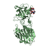
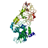
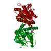
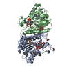
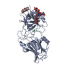
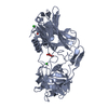
| Entry | Database: PDB / ID: 1af9 | ||||||
|---|---|---|---|---|---|---|---|
| Title | TETANUS NEUROTOXIN C FRAGMENT | ||||||
 Components Components | TETANUS NEUROTOXIN | ||||||
 Keywords Keywords | CLOSTRIDIAL NEUROTOXIN / TETANUS / RECEPTOR BINDING | ||||||
| Function / homology |  Function and homology information Function and homology informationtentoxilysin / symbiont-mediated perturbation of host neurotransmitter secretion / Toxicity of tetanus toxin (tetX) / symbiont-mediated suppression of host exocytosis / transmembrane protein transporter activity / clathrin-coated endocytic vesicle membrane / metalloendopeptidase activity / endocytic vesicle membrane / toxin activity / proteolysis ...tentoxilysin / symbiont-mediated perturbation of host neurotransmitter secretion / Toxicity of tetanus toxin (tetX) / symbiont-mediated suppression of host exocytosis / transmembrane protein transporter activity / clathrin-coated endocytic vesicle membrane / metalloendopeptidase activity / endocytic vesicle membrane / toxin activity / proteolysis / extracellular region / zinc ion binding / plasma membrane / cytosol Similarity search - Function | ||||||
| Biological species |  Clostridium tetani (bacteria) Clostridium tetani (bacteria) | ||||||
| Method |  X-RAY DIFFRACTION / MIRAS / Resolution: 2.7 Å X-RAY DIFFRACTION / MIRAS / Resolution: 2.7 Å | ||||||
 Authors Authors | Umland, T.C. / Wingert, L. / Swaminathan, S. / Furey, W.F. / Schmidt, J.J. / Sax, M. | ||||||
 Citation Citation | Journal: Nat.Struct.Biol. / Year: 1997 Title: Structure of the receptor binding fragment HC of tetanus neurotoxin. Authors: Umland, T.C. / Wingert, L.M. / Swaminathan, S. / Furey, W.F. / Schmidt, J.J. / Sax, M. #1: Journal: Acta Crystallogr.,Sect.D / Year: 1998Title: Crystallization and Preliminary X-Ray Analysis of Tetanus Neurotoxin C Fragment Authors: Umland, T.C. / Wingert, L. / Swaminathan, S. / Schmidt, J.J. / Sax, M. #2: Journal: Thesis, University of Pittsburgh / Year: 1994Title: The Crystal Structures of Human and Rat Clara Cell Phospholipid Binding Proteins and the Preliminary Crystallographic Analysis of Botulinum E Neurotoxin and Tetanus Toxin C-Fragment Authors: Umland, T.C. | ||||||
| History |
|
- Structure visualization
| Structure viewer | Molecule: MolmilJmol/JSmol |
|---|
- Downloads & links
-Download
| PDBx/mmCIF format | 1af9.cif.gz | 102.6 KB | Display | PDBx/mmCIF format |
|---|---|---|---|---|
| PDB format | pdb1af9.ent.gz | 77.2 KB | Display | PDB format |
| PDBx/mmJSON format | 1af9.json.gz | Tree view | PDBx/mmJSON format | |
| Others |  Other downloads Other downloads |
-Validation report
| Arichive directory | https://data.pdbj.org/pub/pdb/validation_reports/af/1af9ftp://data.pdbj.org/pub/pdb/validation_reports/af/1af9 | HTTPS FTP |
|---|
-Related structure data
| Similar structure data |
|---|


-Links
 PDBj
PDBj- Assembly
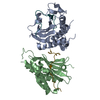
| Deposited unit | 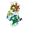
| ||||||||
|---|---|---|---|---|---|---|---|---|---|
| 1 |
| ||||||||
| Unit cell |
|
-Components
| #1: Protein | Mass: 51826.426 Da / Num. of mol.: 1 / Fragment: C FRAGMENT / Mutation: MET INSERTED AT N-TERMINUS Source method: isolated from a genetically manipulated source Source: (gene. exp.) Clostridium tetani (bacteria) / Description: PURCHASED FROM BOEHRINGER MANNHEIM / Production host: |
|---|---|
| #2: Water | ChemComp-HOH /  Mass: 18.015 Da / Num. of mol.: 138 / Source method: isolated from a natural source / Formula: H2O Mass: 18.015 Da / Num. of mol.: 138 / Source method: isolated from a natural source / Formula: H2O |
-Experimental details
-Experiment
| Experiment | Method: X-RAY DIFFRACTION / Number of used crystals: 1 |
|---|
- Sample preparation
| Crystal | Density Matthews: 2.45 Å3/Da / Density % sol: 47 % | |||||||||||||||||||||||||||||||||||
|---|---|---|---|---|---|---|---|---|---|---|---|---|---|---|---|---|---|---|---|---|---|---|---|---|---|---|---|---|---|---|---|---|---|---|---|---|
| Crystal grow | pH: 6.5 Details: CRYSTALLIZATION DROP CONTAINED 3 PARTS RESERVOIR SOLUTION AND 1 PART PROTEIN SOLUTION. RESERVOIR: 14%- 18% PEG 4000, 0.1M IMIDAZOLE, 0.05M MGCL2 AT PH 6.5. PROTEIN SOLUTION: 10MG/ML PROTEIN ...Details: CRYSTALLIZATION DROP CONTAINED 3 PARTS RESERVOIR SOLUTION AND 1 PART PROTEIN SOLUTION. RESERVOIR: 14%- 18% PEG 4000, 0.1M IMIDAZOLE, 0.05M MGCL2 AT PH 6.5. PROTEIN SOLUTION: 10MG/ML PROTEIN AND 0.01M TRIS-HCL AT PH 7.8. PH range: 6.5-7.8 | |||||||||||||||||||||||||||||||||||
| Crystal | *PLUS | |||||||||||||||||||||||||||||||||||
| Crystal grow | *PLUS pH: 7.8 / Method: vapor diffusion, hanging dropDetails: Umland, T.C., (1998) Acta Crystallogr.,Sect.D, 54, 273. | |||||||||||||||||||||||||||||||||||
| Components of the solutions | *PLUS
|
-Data collection
| Diffraction | Mean temperature: 291 K |
|---|---|
| Diffraction source | Source: ROTATING ANODE / Type: RIGAKU RUH2R / Wavelength: 1.5418 |
| Detector | Type: SIEMENS / Detector: AREA DETECTOR / Date: Aug 1, 1994 / Details: SUPPER DOUBLE MIRRORS |
| Radiation | Monochromator: NI FILTER AND SUPPER DOUBLE MIRRORS / Monochromatic (M) / Laue (L): M / Scattering type: x-ray |
| Radiation wavelength | Wavelength: 1.5418 Å / Relative weight: 1 |
| Reflection | Resolution: 2.6→35 Å / Num. obs: 14445 / % possible obs: 91.6 % / Observed criterion σ(I): 1 / Redundancy: 3.3 % / Biso Wilson estimate: 12.9 Å2 / Rmerge(I) obs: 0.066 / Rsym value: 0.076 / Net I/σ(I): 54.8 |
| Reflection shell | Resolution: 2.6→2.7 Å / % possible all: 59.7 |
| Reflection | *PLUS Num. measured all: 47673 |
| Reflection shell | *PLUS % possible obs: 71.2 % |
- Processing
| Software |
| ||||||||||||||||||||||||||||||||||||||||||||||||||||||||||||||||||||||||||||||||
|---|---|---|---|---|---|---|---|---|---|---|---|---|---|---|---|---|---|---|---|---|---|---|---|---|---|---|---|---|---|---|---|---|---|---|---|---|---|---|---|---|---|---|---|---|---|---|---|---|---|---|---|---|---|---|---|---|---|---|---|---|---|---|---|---|---|---|---|---|---|---|---|---|---|---|---|---|---|---|---|---|---|
| Refinement | Method to determine structure: MIRAS / Resolution: 2.7→8 Å / Rfactor Rfree error: 0.0068 / Data cutoff high absF: 100000 / Data cutoff low absF: 0.1 / Isotropic thermal model: RESTRAINED / Cross valid method: THROUGHOUT / σ(F): 2
| ||||||||||||||||||||||||||||||||||||||||||||||||||||||||||||||||||||||||||||||||
| Displacement parameters | Biso mean: 15.6 Å2 | ||||||||||||||||||||||||||||||||||||||||||||||||||||||||||||||||||||||||||||||||
| Refine analyze | Luzzati d res low obs: 8 Å | ||||||||||||||||||||||||||||||||||||||||||||||||||||||||||||||||||||||||||||||||
| Refinement step | Cycle: LAST / Resolution: 2.7→8 Å
| ||||||||||||||||||||||||||||||||||||||||||||||||||||||||||||||||||||||||||||||||
| Refine LS restraints |
| ||||||||||||||||||||||||||||||||||||||||||||||||||||||||||||||||||||||||||||||||
| LS refinement shell | Resolution: 2.7→2.82 Å / Rfactor Rfree error: 0.019 / Total num. of bins used: 8
| ||||||||||||||||||||||||||||||||||||||||||||||||||||||||||||||||||||||||||||||||
| Xplor file |
| ||||||||||||||||||||||||||||||||||||||||||||||||||||||||||||||||||||||||||||||||
| Software | *PLUS Name: X-PLOR / Version: 3.1 / Classification: refinement | ||||||||||||||||||||||||||||||||||||||||||||||||||||||||||||||||||||||||||||||||
| Refine LS restraints | *PLUS
|
