 Movie
Movie Controller


+ Open data

- Basic information
| Entry | Database: PDB / ID: 1af7 | ||||||
|---|---|---|---|---|---|---|---|
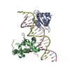
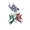
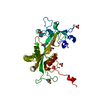
| Title | CHER FROM SALMONELLA TYPHIMURIUM | ||||||
 Components Components | CHEMOTAXIS RECEPTOR METHYLTRANSFERASE CHER | ||||||
 Keywords Keywords | METHYLTRANSFERASE / CHEMOTAXIS RECEPTOR METHYLATION | ||||||
| Function / homology |  Function and homology information Function and homology informationprotein-glutamate O-methyltransferase / protein-glutamate O-methyltransferase activity / methyl accepting chemotaxis protein complex / protein methyltransferase activity / chemotaxis / methylation Similarity search - Function | ||||||
| Biological species |  Salmonella typhimurium (bacteria) Salmonella typhimurium (bacteria) | ||||||
| Method |  X-RAY DIFFRACTION / MIR / Resolution: 2 Å X-RAY DIFFRACTION / MIR / Resolution: 2 Å | ||||||
 Authors Authors | Djordjevic, S. / Stock, A.M. | ||||||
 Citation Citation | Journal: Structure / Year: 1997 Title: Crystal structure of the chemotaxis receptor methyltransferase CheR suggests a conserved structural motif for binding S-adenosylmethionine. Authors: Djordjevic, S. / Stock, A.M. | ||||||
| History |
|
- Structure visualization
| Structure viewer | Molecule: MolmilJmol/JSmol |
|---|
- Downloads & links
-Download
| PDBx/mmCIF format | 1af7.cif.gz | 69.5 KB | Display | PDBx/mmCIF format |
|---|---|---|---|---|
| PDB format | pdb1af7.ent.gz | 51.4 KB | Display | PDB format |
| PDBx/mmJSON format | 1af7.json.gz | Tree view | PDBx/mmJSON format | |
| Others |  Other downloads Other downloads |
-Validation report
| Arichive directory | https://data.pdbj.org/pub/pdb/validation_reports/af/1af7ftp://data.pdbj.org/pub/pdb/validation_reports/af/1af7 | HTTPS FTP |
|---|
-Related structure data
| Similar structure data |
|---|


-Links
 PDBj
PDBj- Assembly
| Deposited unit | 
| ||||||||
|---|---|---|---|---|---|---|---|---|---|
| 1 |
| ||||||||
| Unit cell |
|
-Components
| #1: Protein | Mass: 31533.041 Da / Num. of mol.: 1 Source method: isolated from a genetically manipulated source Details: STRUCTURE INCLUDES S-ADENOSYL-L-HOMOCYSTEINE / Source: (gene. exp.) Salmonella typhimurium (bacteria) / Plasmid: PME43 / Production host: References: UniProt: P07801, protein-glutamate O-methyltransferase |
|---|---|
| #2: Chemical | ChemComp-SAH /   Type: L-peptide linking / Mass: 384.411 Da / Num. of mol.: 1 / Source method: obtained synthetically / Formula: C14H20N6O5S Type: L-peptide linking / Mass: 384.411 Da / Num. of mol.: 1 / Source method: obtained synthetically / Formula: C14H20N6O5S |
| #3: Water | ChemComp-HOH /  Mass: 18.015 Da / Num. of mol.: 111 / Source method: isolated from a natural source / Formula: H2O Mass: 18.015 Da / Num. of mol.: 111 / Source method: isolated from a natural source / Formula: H2O |
-Experimental details
-Experiment
| Experiment | Method: X-RAY DIFFRACTION / Number of used crystals: 1 |
|---|
- Sample preparation
| Crystal | Density Matthews: 2.4 Å3/Da / Density % sol: 40 % | ||||||||||||||||||||
|---|---|---|---|---|---|---|---|---|---|---|---|---|---|---|---|---|---|---|---|---|---|
| Crystal grow | pH: 5.6 Details: MICROSEEDING; 1.2 M AMMONIUM SULFATE, 2% PEG 400, 25 MM SODIUM CITRATE PH 5.6. PRIOR TO DATA COLLECTION CRYSTAL WAS SOAKED IN THE SAME SOLUTION WITH PH ADJUSTED TO 7.0. | ||||||||||||||||||||
| Crystal grow | *PLUS Method: vapor diffusion, hanging drop | ||||||||||||||||||||
| Components of the solutions | *PLUS
|
-Data collection
| Diffraction | Mean temperature: 273 K |
|---|---|
| Diffraction source | Source: ROTATING ANODE / Type: RIGAKU / Wavelength: 1.5418 |
| Detector | Type: RIGAKU RAXIS II / Detector: IMAGE PLATE / Date: Jan 1, 1995 / Details: MIRRORS |
| Radiation | Monochromator: NI FILTER / Monochromatic (M) / Laue (L): M / Scattering type: x-ray |
| Radiation wavelength | Wavelength: 1.5418 Å / Relative weight: 1 |
| Reflection | Resolution: 2.06→34.3 Å / Num. obs: 18331 / % possible obs: 93 % / Observed criterion σ(I): 2 / Redundancy: 2.1 % / Rmerge(I) obs: 0.049 / Net I/σ(I): 11 |
| Reflection shell | Resolution: 2.06→2.25 Å / Redundancy: 2 % / Rmerge(I) obs: 0.013 / Mean I/σ(I) obs: 6.4 / % possible all: 91.8 |
- Processing
| Software |
| ||||||||||||||||
|---|---|---|---|---|---|---|---|---|---|---|---|---|---|---|---|---|---|
| Refinement | Method to determine structure: MIR / Resolution: 2→34.3 Å / σ(F): 2 Details: RESIDUES 13 - 22, AND 184 - 194 BELONG TO WEAKLY DEFINED REGIONS. RESIDUE SER 125 HAS PHI PSI VALUES OUTSIDE OF THE EXPECTED RANGE BUT THIS IS REAL BUT THIS IS REAL AND IT IS TRUE FOR ALL ...Details: RESIDUES 13 - 22, AND 184 - 194 BELONG TO WEAKLY DEFINED REGIONS. RESIDUE SER 125 HAS PHI PSI VALUES OUTSIDE OF THE EXPECTED RANGE BUT THIS IS REAL BUT THIS IS REAL AND IT IS TRUE FOR ALL METHYLTRANSFERASES IN THAT POSITION. THAT RESIDUE IS PART OF THE LOOP THAT IS FOUND TO BE IMPORTANT FOR THE CATALYTIC PART OF THE LOOP THAT IS FOUND TO BE IMPORTANT FOR THE CATALYTIC ACTIVITY AND UNIQUE FOR METHYLTRANSFERASES. BACKBONE ANGLES FOR REGION 121 - 127 ARE ALSO DEVIATING BUT THEY TOO ARE INVOLVED IN FORMATION OF THIS SPECIFIC LOOP.
| ||||||||||||||||
| Refinement step | Cycle: LAST / Resolution: 2→34.3 Å
| ||||||||||||||||
| Software | *PLUS Name: ARP / Classification: refinement | ||||||||||||||||
| Refinement | *PLUS Num. reflection all: 18331 / Rfactor obs: 0.196 | ||||||||||||||||
| Solvent computation | *PLUS | ||||||||||||||||
| Displacement parameters | *PLUS | ||||||||||||||||
| Refine LS restraints | *PLUS
|
