 Movie
Movie Controller

[English] 日本語
 Yorodumi
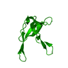
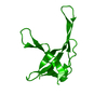
- PDB-1ae3: MUTANT R82C OF GENE V PROTEIN (SINGLE-STRANDED DNA BINDING PROTEIN) -
+ Open data

- Basic information
| Entry | Database: PDB / ID: 1ae3 | ||||||
|---|---|---|---|---|---|---|---|
| Title | MUTANT R82C OF GENE V PROTEIN (SINGLE-STRANDED DNA BINDING PROTEIN) | ||||||
 Components Components | GENE V PROTEIN | ||||||
 Keywords Keywords | DNA BINDING PROTEIN / MUTANT / R82C / GVP / SINGLE-STRANDED DNA BINDING PROTEIN / DNA REPLICATION / DNA-BINDING PROTEIN | ||||||
| Function / homology |  Function and homology information Function and homology informationrolling circle single-stranded viral DNA replication / single-stranded DNA binding / DNA replication Similarity search - Function | ||||||
| Biological species |  | ||||||
| Method |  X-RAY DIFFRACTION / DIFFERENCE FOURIERS / Resolution: 2 Å X-RAY DIFFRACTION / DIFFERENCE FOURIERS / Resolution: 2 Å | ||||||
 Authors Authors | Su, S. / Gao, Y.-G. / Zhang, H. / Terwilliger, T.C. / Wang, A.H.-J. | ||||||
 Citation Citation | Journal: Protein Sci. / Year: 1997 Title: Analyses of the stability and function of three surface mutants (R82C, K69H, and L32R) of the gene V protein from Ff phage by X-ray crystallography. Authors: Su, S. / Gao, Y.G. / Zhang, H. / Terwilliger, T.C. / Wang, A.H. | ||||||
| History |
|
- Structure visualization
| Structure viewer | Molecule: MolmilJmol/JSmol |
|---|
- Downloads & links
-Download
| PDBx/mmCIF format | 1ae3.cif.gz | 34.1 KB | Display | PDBx/mmCIF format |
|---|---|---|---|---|
| PDB format | pdb1ae3.ent.gz | 23.3 KB | Display | PDB format |
| PDBx/mmJSON format | 1ae3.json.gz | Tree view | PDBx/mmJSON format | |
| Others |  Other downloads Other downloads |
-Validation report
| Summary document | 1ae3_validation.pdf.gz | 410 KB | Display | wwPDB validaton report |
|---|---|---|---|---|
| Full document | 1ae3_full_validation.pdf.gz | 411.6 KB | Display | |
| Data in XML | 1ae3_validation.xml.gz | 6.4 KB | Display | |
| Data in CIF | 1ae3_validation.cif.gz | 7.8 KB | Display | |
| Arichive directory | https://data.pdbj.org/pub/pdb/validation_reports/ae/1ae3ftp://data.pdbj.org/pub/pdb/validation_reports/ae/1ae3 | HTTPS FTP |
-Related structure data


-Links
 PDBj
PDBj- Assembly
| Deposited unit | 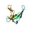
| ||||||||
|---|---|---|---|---|---|---|---|---|---|
| 1 | 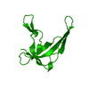
| ||||||||
| Unit cell |
|
-Components
| #1: Protein | Mass: 9515.983 Da / Num. of mol.: 1 / Mutation: R82C Source method: isolated from a genetically manipulated source Source: (gene. exp.) |
|---|---|
| #2: Water | ChemComp-HOH /  Mass: 18.015 Da / Num. of mol.: 47 / Source method: isolated from a natural source / Formula: H2O Mass: 18.015 Da / Num. of mol.: 47 / Source method: isolated from a natural source / Formula: H2O |
-Experimental details
-Experiment
| Experiment | Method: X-RAY DIFFRACTION / Number of used crystals: 1 |
|---|
- Sample preparation
| Crystal | Density Matthews: 2.2 Å3/Da / Density % sol: 44 % | |||||||||||||||||||||||||
|---|---|---|---|---|---|---|---|---|---|---|---|---|---|---|---|---|---|---|---|---|---|---|---|---|---|---|
| Crystal grow | pH: 7 Details: R82C GVP WAS CRYSTALLIZED FROM SOLUTIONS CONTAINING 10 MG/ML PROTEIN, 35 MM TRIS (PH7.0), AND 4.7% PEG 4000 (W/V), EQUILIBRATED AGAINST 6% PEG 4000. | |||||||||||||||||||||||||
| Crystal grow | *PLUS Method: vapor diffusion | |||||||||||||||||||||||||
| Components of the solutions | *PLUS
|
-Data collection
| Diffraction | Mean temperature: 285 K |
|---|---|
| Diffraction source | Source: ROTATING ANODE / Type: RIGAKU RUH2R / Wavelength: 1.5418 |
| Detector | Type: RIGAKU / Detector: IMAGE PLATE / Date: Jan 1, 1996 / Details: COLLIMATORS |
| Radiation | Monochromator: GRAPHITE(002) / Monochromatic (M) / Laue (L): M / Scattering type: x-ray |
| Radiation wavelength | Wavelength: 1.5418 Å / Relative weight: 1 |
| Reflection | Resolution: 2→15 Å / Num. obs: 4758 / % possible obs: 79.2 % / Observed criterion σ(I): 2 / Biso Wilson estimate: 20.8 Å2 / Rmerge(I) obs: 0.088 / Net I/σ(I): 7.2 |
| Reflection shell | Resolution: 2→2.09 Å / % possible all: 58.8 |
| Reflection shell | *PLUS % possible obs: 58.8 % |
- Processing
| Software |
| ||||||||||||||||||||||||||||||||||||||||||||||||||||||||||||||||||||||||||||||||
|---|---|---|---|---|---|---|---|---|---|---|---|---|---|---|---|---|---|---|---|---|---|---|---|---|---|---|---|---|---|---|---|---|---|---|---|---|---|---|---|---|---|---|---|---|---|---|---|---|---|---|---|---|---|---|---|---|---|---|---|---|---|---|---|---|---|---|---|---|---|---|---|---|---|---|---|---|---|---|---|---|---|
| Refinement | Method to determine structure: DIFFERENCE FOURIERS / Resolution: 2→8 Å / Rfactor Rfree error: 0.013 / Data cutoff high absF: 100000 / Data cutoff low absF: 0 / Isotropic thermal model: RESTRAINED / Cross valid method: THROUGHOUT / σ(F): 2
| ||||||||||||||||||||||||||||||||||||||||||||||||||||||||||||||||||||||||||||||||
| Displacement parameters | Biso mean: 26.1 Å2
| ||||||||||||||||||||||||||||||||||||||||||||||||||||||||||||||||||||||||||||||||
| Refine analyze |
| ||||||||||||||||||||||||||||||||||||||||||||||||||||||||||||||||||||||||||||||||
| Refinement step | Cycle: LAST / Resolution: 2→8 Å
| ||||||||||||||||||||||||||||||||||||||||||||||||||||||||||||||||||||||||||||||||
| Refine LS restraints |
| ||||||||||||||||||||||||||||||||||||||||||||||||||||||||||||||||||||||||||||||||
| LS refinement shell | Resolution: 2→2.09 Å / Rfactor Rfree error: 0.043 / Total num. of bins used: 8
| ||||||||||||||||||||||||||||||||||||||||||||||||||||||||||||||||||||||||||||||||
| Xplor file |
| ||||||||||||||||||||||||||||||||||||||||||||||||||||||||||||||||||||||||||||||||
| Software | *PLUS Name: X-PLOR / Version: 3.1 / Classification: refinement | ||||||||||||||||||||||||||||||||||||||||||||||||||||||||||||||||||||||||||||||||
| Refinement | *PLUS | ||||||||||||||||||||||||||||||||||||||||||||||||||||||||||||||||||||||||||||||||
| Solvent computation | *PLUS | ||||||||||||||||||||||||||||||||||||||||||||||||||||||||||||||||||||||||||||||||
| Displacement parameters | *PLUS | ||||||||||||||||||||||||||||||||||||||||||||||||||||||||||||||||||||||||||||||||
| Refine LS restraints | *PLUS
| ||||||||||||||||||||||||||||||||||||||||||||||||||||||||||||||||||||||||||||||||
| LS refinement shell | *PLUS Rfactor obs: 0.226 |
