 Movie
Movie Controller

[English] 日本語
 Yorodumi
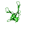
- PDB-1vqe: GENE V PROTEIN MUTANT WITH VAL 35 REPLACED BY ILE 35 AND ILE 47 R... -
+ Open data

- Basic information
| Entry | Database: PDB / ID: 1vqe | ||||||
|---|---|---|---|---|---|---|---|
| Title | GENE V PROTEIN MUTANT WITH VAL 35 REPLACED BY ILE 35 AND ILE 47 REPLACED BY MET 47 (V35I, I47M) | ||||||
 Components Components | GENE V PROTEIN | ||||||
 Keywords Keywords | DNA BINDING PROTEIN / DNA-BINDING PROTEIN / GENE V / MUTANT | ||||||
| Function / homology |  Function and homology information Function and homology informationrolling circle single-stranded viral DNA replication / single-stranded DNA binding / DNA replication Similarity search - Function | ||||||
| Biological species |  Enterobacteria phage f1 (virus) Enterobacteria phage f1 (virus) | ||||||
| Method |  X-RAY DIFFRACTION / Resolution: 1.8 Å X-RAY DIFFRACTION / Resolution: 1.8 Å | ||||||
 Authors Authors | Skinner, M.M. / Terwilliger, T.C. | ||||||
 Citation Citation | Journal: Proc.Natl.Acad.Sci.USA / Year: 1996 Title: Potential use of additivity of mutational effects in simplifying protein engineering. Authors: Skinner, M.M. / Terwilliger, T.C. #1: Journal: Acta Crystallogr.,Sect.D / Year: 1995Title: Difference Refinement: Obtaining Differences between Two Related Structures Authors: Terwilliger, T.C. / Berendzen, J. #2: Journal: Proc.Natl.Acad.Sci.USA / Year: 1994Title: Structure of the Gene V Protein of Bacteriophage F1 Determined by Multiwavelength X-Ray Diffraction on the Selenomethionyl Protein Authors: Skinner, M.M. / Zhang, H. / Leschnitzer, D.H. / Guan, Y. / Bellamy, H. / Sweet, R.M. / Gray, C.W. / Konings, R.N. / Wang, A.H. / Terwilliger, T.C. | ||||||
| History |
|
- Structure visualization
| Structure viewer | Molecule: MolmilJmol/JSmol |
|---|
- Downloads & links
-Download
| PDBx/mmCIF format | 1vqe.cif.gz | 32.2 KB | Display | PDBx/mmCIF format |
|---|---|---|---|---|
| PDB format | pdb1vqe.ent.gz | 21.5 KB | Display | PDB format |
| PDBx/mmJSON format | 1vqe.json.gz | Tree view | PDBx/mmJSON format | |
| Others |  Other downloads Other downloads |
-Validation report
| Arichive directory | https://data.pdbj.org/pub/pdb/validation_reports/vq/1vqeftp://data.pdbj.org/pub/pdb/validation_reports/vq/1vqe | HTTPS FTP |
|---|
-Related structure data


-Links
 PDBj
PDBj- Assembly
| Deposited unit | 
| ||||||||
|---|---|---|---|---|---|---|---|---|---|
| 1 | 
| ||||||||
| Unit cell |
|
-Components
| #1: Protein | Mass: 9731.279 Da / Num. of mol.: 1 / Mutation: V35I, I47M Source method: isolated from a genetically manipulated source Source: (gene. exp.) Enterobacteria phage f1 (virus) / Genus: Inovirus / Species: Enterobacteria phage M13 / Production host:  |
|---|---|
| #2: Water | ChemComp-HOH /  Mass: 18.015 Da / Num. of mol.: 44 / Source method: isolated from a natural source / Formula: H2O Mass: 18.015 Da / Num. of mol.: 44 / Source method: isolated from a natural source / Formula: H2O |
-Experimental details
-Experiment
| Experiment | Method: X-RAY DIFFRACTION |
|---|
- Sample preparation
| Crystal | Density Matthews: 2.27 Å3/Da / Density % sol: 45 % | |||||||||||||||||||||||||
|---|---|---|---|---|---|---|---|---|---|---|---|---|---|---|---|---|---|---|---|---|---|---|---|---|---|---|
| Crystal grow | *PLUS pH: 7.6 / Method: vapor diffusionDetails: Skinner, M.M., (1994) Proc.Nat.Acad.Sci.USA, 91, 2071. | |||||||||||||||||||||||||
| Components of the solutions | *PLUS
|
-Data collection
| Diffraction source | Wavelength: 1.5418 |
|---|---|
| Detector | Type: MARRESEARCH / Detector: IMAGE PLATE / Date: May 1, 1994 |
| Radiation | Monochromatic (M) / Laue (L): M / Scattering type: x-ray |
| Radiation wavelength | Wavelength: 1.5418 Å / Relative weight: 1 |
| Reflection | Num. obs: 8006 / % possible obs: 97.3 % / Redundancy: 4 % / Rmerge(I) obs: 0.043 |
| Reflection | *PLUS Highest resolution: 1.8 Å |
- Processing
| Software |
| ||||||||||||||||||||||||||||||||||||||||||||||||||||||||||||
|---|---|---|---|---|---|---|---|---|---|---|---|---|---|---|---|---|---|---|---|---|---|---|---|---|---|---|---|---|---|---|---|---|---|---|---|---|---|---|---|---|---|---|---|---|---|---|---|---|---|---|---|---|---|---|---|---|---|---|---|---|---|
| Refinement | Resolution: 1.8→5 Å / σ(F): 2 Details: SIDE CHAIN DISORDERED DENSITY FOR GLN 12 IS MODELED STEREOCHEMICALLY.
| ||||||||||||||||||||||||||||||||||||||||||||||||||||||||||||
| Displacement parameters | Biso mean: 20 Å2 | ||||||||||||||||||||||||||||||||||||||||||||||||||||||||||||
| Refine analyze | Luzzati coordinate error obs: 0.22 Å | ||||||||||||||||||||||||||||||||||||||||||||||||||||||||||||
| Refinement step | Cycle: LAST / Resolution: 1.8→5 Å
| ||||||||||||||||||||||||||||||||||||||||||||||||||||||||||||
| Refine LS restraints |
|
