 Movie
Movie Controller

[English] 日本語
 Yorodumi
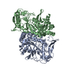
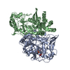
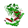
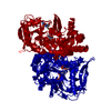
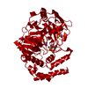
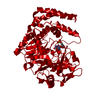
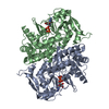
- PDB-1ade: STRUCTURE OF ADENYLOSUCCINATE SYNTHETASE PH 7 AT 25 DEGREES CELSIUS -
+ Open data

- Basic information
| Entry | Database: PDB / ID: 1ade | ||||||
|---|---|---|---|---|---|---|---|
| Title | STRUCTURE OF ADENYLOSUCCINATE SYNTHETASE PH 7 AT 25 DEGREES CELSIUS | ||||||
 Components Components | ADENYLOSUCCINATE SYNTHETASE | ||||||
 Keywords Keywords | LIGASE (SYNTHETASE) / PURINE NUCLEOTIDE BIOSYNTHESIS / LIGASE / GTP-HYDROLYZING ENZYMES | ||||||
| Function / homology |  Function and homology information Function and homology informationadenylosuccinate synthase / adenylosuccinate synthase activity / adenosine biosynthetic process / IMP metabolic process / 'de novo' AMP biosynthetic process / nucleobase-containing small molecule interconversion / purine nucleotide biosynthetic process / guanosine tetraphosphate binding / DNA damage response / GTP binding ...adenylosuccinate synthase / adenylosuccinate synthase activity / adenosine biosynthetic process / IMP metabolic process / 'de novo' AMP biosynthetic process / nucleobase-containing small molecule interconversion / purine nucleotide biosynthetic process / guanosine tetraphosphate binding / DNA damage response / GTP binding / magnesium ion binding / membrane / cytoplasm / cytosol Similarity search - Function | ||||||
| Biological species |  | ||||||
| Method |  X-RAY DIFFRACTION / Resolution: 2 Å X-RAY DIFFRACTION / Resolution: 2 Å | ||||||
 Authors Authors | Silva, M.M. / Poland, B.W. / Hoffman, C.M. / Fromm, H.J. / Honzatko, R.B. | ||||||
 Citation Citation | Journal: J.Mol.Biol. / Year: 1995 Title: Refined crystal structures of unligated adenylosuccinate synthetase from Escherichia coli. Authors: Silva, M.M. / Poland, B.W. / Hoffman, C.R. / Fromm, H.J. / Honzatko, R.B. #1: Journal: J.Biol.Chem. / Year: 1993Title: Crystal Structure of Adenylosuccinate Synthetase from Escherichia Coli Authors: Poland, B.W. / Silva, M.M. / Serra, M.A. / Cho, Y. / Kim, K.H. / Harris, E.M.S. / Honzatko, R.B. | ||||||
| History |
|
- Structure visualization
| Structure viewer | Molecule: MolmilJmol/JSmol |
|---|
- Downloads & links
-Download
| PDBx/mmCIF format | 1ade.cif.gz | 239 KB | Display | PDBx/mmCIF format |
|---|---|---|---|---|
| PDB format | pdb1ade.ent.gz | 193.8 KB | Display | PDB format |
| PDBx/mmJSON format | 1ade.json.gz | Tree view | PDBx/mmJSON format | |
| Others |  Other downloads Other downloads |
-Validation report
| Arichive directory | https://data.pdbj.org/pub/pdb/validation_reports/ad/1adeftp://data.pdbj.org/pub/pdb/validation_reports/ad/1ade | HTTPS FTP |
|---|
-Related structure data


-Links
 PDBj
PDBj- Assembly
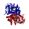
| Deposited unit | 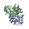
| ||||||||
|---|---|---|---|---|---|---|---|---|---|
| 1 |
| ||||||||
| Unit cell |
| ||||||||
| Atom site foot note | 1: CIS PROLINE - PRO A 236 / 2: CIS PROLINE - PRO B 236 | ||||||||
| Noncrystallographic symmetry (NCS) | NCS oper: (Code: given Matrix: (0.54489, -0.21018, -0.81174), Vector: Details | MTRIX THE TRANSFORMATIONS PRESENTED ON MTRIX RECORDS BELOW DESCRIBE NON-CRYSTALLOGRAPHIC RELATIONSHIPS AMONG THE VARIOUS DOMAINS IN THIS ENTRY. APPLYING THE APPROPRIATE MTRIX TRANSFORMATION TO THE RESIDUES LISTED FIRST WILL YIELD APPROXIMATE COORDINATES FOR THE RESIDUES LISTED SECOND. APPLIED TO TRANSFORMED TO MTRIX RESIDUES RESIDUES RMSD M1 B 1 .. B 431 A 1 .. A 431 1.374 | |
-Components
| #1: Protein | Mass: 47269.598 Da / Num. of mol.: 2 / Source method: isolated from a natural source Details: COLI GENETIC STOCK CENTER, STRAIN NUMBER 5408. GIFT FROM DR. B. BACHMAN, GENETIC CENTER, YALE UNIVERSITY Source: (natural) #2: Water | ChemComp-HOH / |  Mass: 18.015 Da / Num. of mol.: 574 / Source method: isolated from a natural source / Formula: H2O Mass: 18.015 Da / Num. of mol.: 574 / Source method: isolated from a natural source / Formula: H2OCompound details | TURN TURN_ID: T4, TYPE I (ONE OR MORE OF THE PHI, PSI ANGLES DEVIATE BY MORE THAN PLUS, MINUS 45 ...TURN TURN_ID: T4, TYPE I (ONE OR MORE OF THE PHI, PSI ANGLES DEVIATE BY MORE THAN PLUS, MINUS 45 DEGREE FROM THE IDEAL VALUES USED BY WILMOT & THORNTON(1989)). TURN_ID: T10, TYPE I (ONE OR MORE OF THE PHI, PSI ANGLES DEVIATE BY MORE THAN PLUS, MINUS 45 DEGREE FROM THE IDEAL VALUES USED BY WILMOT & THORNTON(1989)). TURN_ID: T16, TYPE VIII (ONE OR MORE OF THE PHI, PSI ANGLES DEVIATE BY MORE THAN PLUS,MINUS 45 DEGREE FROM THE IDEAL VALUES USED BY WILMOT & THORNTON(1989)). TURN_ID: T28, TYPE I (ONE OR MORE OF THE PHI, PSI ANGLES DEVIATE BY MORE THAN PLUS, MINUS 45 DEGREE FROM THE IDEAL VALUES USED BY WILMOT & THORNTON(1989)). TURN_ID: T29, TYPE I (ONE OR MORE OF THE PHI, PSI ANGLES DEVIATE BY MORE THAN PLUS, MINUS 45 DEGREE FROM THE IDEAL VALUES USED BY WILMOT & THORNTON(1989)). TURN_ID: T4, TYPE I (ONE OR MORE OF THE PHI, PSI ANGLES DEVIATE BY MORE THAN PLUS, MINUS 45 DEGREE FROM THE IDEAL VALUES USED BY WILMOT & THORNTON(1989)). TURN_ID: T10, TYPE I (ONE OR MORE OF THE PHI, PSI ANGLES DEVIATE BY MORE THAN PLUS, MINUS 45 DEGREE FROM THE IDEAL VALUES USED BY WILMOT & THORNTON(1989)). TURN_ID: T16, TYPE VIII (ONE OR MORE OF THE PHI, PSI ANGLES DEVIATE BY MORE THAN PLUS,MINUS 45 DEGREE FROM THE IDEAL VALUES USED BY WILMOT & THORNTON(1989)). TURN_ID: T28, TYPE I (ONE OR MORE OF THE PHI, PSI ANGLES DEVIATE BY MORE THAN PLUS, MINUS 45 DEGREE FROM THE IDEAL VALUES USED BY WILMOT & THORNTON(1989)). TURN_ID: T29, TYPE I (ONE OR MORE OF THE PHI, PSI ANGLES DEVIATE BY MORE THAN PLUS, MINUS 45 DEGREE FROM THE IDEAL VALUES USED BY WILMOT & THORNTON(1989)). | |
|---|
-Experimental details
-Experiment
| Experiment | Method: X-RAY DIFFRACTION |
|---|
- Sample preparation
| Crystal | Density Matthews: 2.2 Å3/Da / Density % sol: 44.14 % | ||||||||||||||||||||||||||||||||||||
|---|---|---|---|---|---|---|---|---|---|---|---|---|---|---|---|---|---|---|---|---|---|---|---|---|---|---|---|---|---|---|---|---|---|---|---|---|---|
| Crystal grow | pH: 7 / Details: pH 7.0 | ||||||||||||||||||||||||||||||||||||
| Crystal grow | *PLUS Temperature: 20-25 ℃ / Method: vapor diffusion, hanging drop | ||||||||||||||||||||||||||||||||||||
| Components of the solutions | *PLUS
|
-Data collection
| Diffraction | Mean temperature: 298 K |
|---|---|
| Diffraction source | Wavelength: 1.5418 Å |
| Detector | Type: XUONG-HAMLIN MULTIWIRE / Detector: AREA DETECTOR |
| Radiation | Monochromatic (M) / Laue (L): M / Scattering type: x-ray |
| Radiation wavelength | Wavelength: 1.5418 Å / Relative weight: 1 |
| Reflection | Num. obs: 32567 / % possible obs: 98 % / Redundancy: 3.2 % / Rmerge(I) obs: 0.068 |
| Reflection | *PLUS Highest resolution: 2 Å / Num. obs: 54490 / Num. measured all: 174373 / Rmerge(I) obs: 0.068 |
| Reflection shell | *PLUS Highest resolution: 2 Å / Lowest resolution: 2.07 Å / % possible obs: 95 % |
- Processing
| Software |
| ||||||||||||||||||||||||||||||||||||||||||||||||||||||||||||
|---|---|---|---|---|---|---|---|---|---|---|---|---|---|---|---|---|---|---|---|---|---|---|---|---|---|---|---|---|---|---|---|---|---|---|---|---|---|---|---|---|---|---|---|---|---|---|---|---|---|---|---|---|---|---|---|---|---|---|---|---|---|
| Refinement | Resolution: 2→5 Å /
| ||||||||||||||||||||||||||||||||||||||||||||||||||||||||||||
| Displacement parameters | Biso mean: 27 Å2 | ||||||||||||||||||||||||||||||||||||||||||||||||||||||||||||
| Refine analyze | Luzzati coordinate error obs: 0.25 Å | ||||||||||||||||||||||||||||||||||||||||||||||||||||||||||||
| Refinement step | Cycle: LAST / Resolution: 2→5 Å
| ||||||||||||||||||||||||||||||||||||||||||||||||||||||||||||
| Refine LS restraints |
| ||||||||||||||||||||||||||||||||||||||||||||||||||||||||||||
| Refinement | *PLUS Num. reflection obs: 49130 | ||||||||||||||||||||||||||||||||||||||||||||||||||||||||||||
| Solvent computation | *PLUS | ||||||||||||||||||||||||||||||||||||||||||||||||||||||||||||
| Displacement parameters | *PLUS | ||||||||||||||||||||||||||||||||||||||||||||||||||||||||||||
| Refine LS restraints | *PLUS
|
