 Movie
Movie Controller


+ Open data

- Basic information
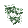
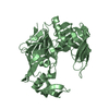
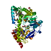
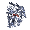
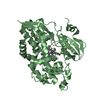
| Entry | Database: PDB / ID: 1ab8 | ||||||
|---|---|---|---|---|---|---|---|
| Title | RAT TYPE II ADENYLYL CYCLASE C2 DOMAIN/FORSKOLIN COMPLEX | ||||||
 Components Components | ADENYLYL CYCLASE | ||||||
 Keywords Keywords | LYASE / ADENYLYL CYCLASE / PLASMID / COMPLEX (TRANSFERASE-INHIBITOR) | ||||||
| Function / homology |  Function and homology information Function and homology informationAdenylate cyclase activating pathway / PKA activation / Hedgehog 'off' state / Adenylate cyclase inhibitory pathway / adenylate cyclase / adenylate cyclase activity / G alpha (z) signalling events / cAMP biosynthetic process / adenylate cyclase binding / cellular response to forskolin ...Adenylate cyclase activating pathway / PKA activation / Hedgehog 'off' state / Adenylate cyclase inhibitory pathway / adenylate cyclase / adenylate cyclase activity / G alpha (z) signalling events / cAMP biosynthetic process / adenylate cyclase binding / cellular response to forskolin / G-protein beta/gamma-subunit complex binding / adenylate cyclase-modulating G protein-coupled receptor signaling pathway / manganese ion binding / adenylate cyclase-activating G protein-coupled receptor signaling pathway / intracellular signal transduction / membrane raft / dendrite / magnesium ion binding / protein-containing complex / ATP binding / membrane / plasma membrane / cytoplasm Similarity search - Function | ||||||
| Biological species |  | ||||||
| Method |  X-RAY DIFFRACTION / SYNCHROTRON / MIRAS / Resolution: 2.2 Å X-RAY DIFFRACTION / SYNCHROTRON / MIRAS / Resolution: 2.2 Å | ||||||
 Authors Authors | Zhang, G. / Hurley, J.H. | ||||||
 Citation Citation | Journal: Nature / Year: 1997 Title: Structure of the adenylyl cyclase catalytic core. Authors: Zhang, G. / Liu, Y. / Ruoho, A.E. / Hurley, J.H. | ||||||
| History |
|
- Structure visualization
| Structure viewer | Molecule: MolmilJmol/JSmol |
|---|
- Downloads & links
-Download
| PDBx/mmCIF format | 1ab8.cif.gz | 86.6 KB | Display | PDBx/mmCIF format |
|---|---|---|---|---|
| PDB format | pdb1ab8.ent.gz | 65.1 KB | Display | PDB format |
| PDBx/mmJSON format | 1ab8.json.gz | Tree view | PDBx/mmJSON format | |
| Others |  Other downloads Other downloads |
-Validation report
| Arichive directory | https://data.pdbj.org/pub/pdb/validation_reports/ab/1ab8ftp://data.pdbj.org/pub/pdb/validation_reports/ab/1ab8 | HTTPS FTP |
|---|
-Related structure data
| Similar structure data |
|---|


-Links
 PDBj
PDBj


- Assembly
| Deposited unit | 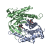
| ||||||||
|---|---|---|---|---|---|---|---|---|---|
| 1 |
| ||||||||
| 2 | 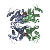
| ||||||||
| Unit cell |
| ||||||||
| Components on special symmetry positions |
| ||||||||
| Noncrystallographic symmetry (NCS) | NCS oper: (Code: given Matrix: (-0.198769, -0.978716, -0.051049), Vector: |
-Components
| #1: Protein | Mass: 24447.781 Da / Num. of mol.: 2 / Fragment: C2 DOMAIN Source method: isolated from a genetically manipulated source Source: (gene. exp.)  #2: Chemical |   Mass: 410.501 Da / Num. of mol.: 2 / Source method: obtained synthetically / Formula: C22H34O7 Mass: 410.501 Da / Num. of mol.: 2 / Source method: obtained synthetically / Formula: C22H34O7#3: Water | ChemComp-HOH / |  Mass: 18.015 Da / Num. of mol.: 113 / Source method: isolated from a natural source / Formula: H2O Mass: 18.015 Da / Num. of mol.: 113 / Source method: isolated from a natural source / Formula: H2O |
|---|
-Experimental details
-Experiment
| Experiment | Method: X-RAY DIFFRACTION / Number of used crystals: 1 |
|---|
- Sample preparation
| Crystal | Density Matthews: 3 Å3/Da / Density % sol: 55 % | ||||||||||||||||||||||||||||||||||||||||||||||||||||||
|---|---|---|---|---|---|---|---|---|---|---|---|---|---|---|---|---|---|---|---|---|---|---|---|---|---|---|---|---|---|---|---|---|---|---|---|---|---|---|---|---|---|---|---|---|---|---|---|---|---|---|---|---|---|---|---|
| Crystal grow | pH: 5.9 Details: 1.1 M AM SO4, 0.1 M PHOSPHATE, PH 5.9, 2 % DMSO, 1 MM FORSKOLIN, 10 MM DTT, 80 MM NACL, 50 MM TRIS HCL | ||||||||||||||||||||||||||||||||||||||||||||||||||||||
| Crystal grow | *PLUS Temperature: 21-24 ℃ / Method: unknown / PH range low: 5.9 / PH range high: 5.8 | ||||||||||||||||||||||||||||||||||||||||||||||||||||||
| Components of the solutions | *PLUS
|
-Data collection
| Diffraction | Mean temperature: 95 K |
|---|---|
| Diffraction source | Source: SYNCHROTRON / Site: CHESS  / Beamline: A1 / Wavelength: 0.908 / Beamline: A1 / Wavelength: 0.908 |
| Detector | Type: FUJI / Detector: IMAGE PLATE / Date: Dec 1, 1996 |
| Radiation | Monochromator: SI(111) / Monochromatic (M) / Laue (L): M / Scattering type: x-ray |
| Radiation wavelength | Wavelength: 0.908 Å / Relative weight: 1 |
| Reflection | Resolution: 2.2→40 Å / Num. obs: 17326 / % possible obs: 91 % / Observed criterion σ(I): 0 / Redundancy: 3.5 % / Rmerge(I) obs: 0.067 / Rsym value: 0.067 / Net I/σ(I): 15 |
| Reflection shell | Resolution: 2.2→2.28 Å / Redundancy: 1 % / Mean I/σ(I) obs: 3 / % possible all: 40 |
| Reflection | *PLUS Num. measured all: 51618 |
- Processing
| Software |
| ||||||||||||||||||||||||||||||||||||||||||||||||||||||||||||
|---|---|---|---|---|---|---|---|---|---|---|---|---|---|---|---|---|---|---|---|---|---|---|---|---|---|---|---|---|---|---|---|---|---|---|---|---|---|---|---|---|---|---|---|---|---|---|---|---|---|---|---|---|---|---|---|---|---|---|---|---|---|
| Refinement | Method to determine structure: MIRAS / Resolution: 2.2→6 Å / Data cutoff high absF: 10000000 / Data cutoff low absF: 1.0E-5 / Isotropic thermal model: RESTRAINED / Cross valid method: FREE R / σ(F): 2 / Details: ANISOTROPIC DIFFRACTION
| ||||||||||||||||||||||||||||||||||||||||||||||||||||||||||||
| Displacement parameters | Biso mean: 41 Å2 | ||||||||||||||||||||||||||||||||||||||||||||||||||||||||||||
| Refine analyze | Luzzati d res low obs: 6 Å | ||||||||||||||||||||||||||||||||||||||||||||||||||||||||||||
| Refinement step | Cycle: LAST / Resolution: 2.2→6 Å
| ||||||||||||||||||||||||||||||||||||||||||||||||||||||||||||
| Refine LS restraints |
| ||||||||||||||||||||||||||||||||||||||||||||||||||||||||||||
| Refine LS restraints NCS | NCS model details: RESTRAINED | ||||||||||||||||||||||||||||||||||||||||||||||||||||||||||||
| Xplor file |
| ||||||||||||||||||||||||||||||||||||||||||||||||||||||||||||
| Software | *PLUS Name: X-PLOR / Version: 3.8 / Classification: refinement | ||||||||||||||||||||||||||||||||||||||||||||||||||||||||||||
| Refine LS restraints | *PLUS
|
