 Movie
Movie Controller

[English] 日本語
 Yorodumi
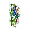
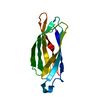
- PDB-1a7w: CRYSTAL STRUCTURE OF THE HISTONE HMFB FROM METHANOTHERMUS FERVIDUS -
+ Open data

- Basic information
| Entry | Database: PDB / ID: 1a7w | ||||||
|---|---|---|---|---|---|---|---|
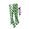
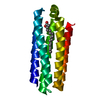
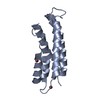
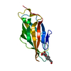
| Title | CRYSTAL STRUCTURE OF THE HISTONE HMFB FROM METHANOTHERMUS FERVIDUS | ||||||
 Components Components | HISTONE HMFB | ||||||
 Keywords Keywords | HISTONE | ||||||
| Function / homology |  Function and homology information Function and homology informationDNA topological change / protein homooligomerization / chromosome / double-stranded DNA binding / protein heterodimerization activity / protein homodimerization activity / cytoplasm Similarity search - Function | ||||||
| Biological species |   Methanothermus fervidus (archaea) Methanothermus fervidus (archaea) | ||||||
| Method |  X-RAY DIFFRACTION / MOLECULAR REPLACEMENT / Resolution: 1.55 Å X-RAY DIFFRACTION / MOLECULAR REPLACEMENT / Resolution: 1.55 Å | ||||||
 Authors Authors | Decanniere, K. / Sandman, K. / Reeve, J.N. / Heinemann, U. | ||||||
 Citation Citation | Journal: J.Mol.Biol. / Year: 2000 Title: Crystal structures of recombinant histones HMfA and HMfB from the hyperthermophilic archaeon Methanothermus fervidus. Authors: Decanniere, K. / Babu, A.M. / Sandman, K. / Reeve, J.N. / Heinemann, U. #1: Journal: Proteins / Year: 1996Title: Crystallization and Preliminary X-Ray Characterization of the Methanothermus Fervidus Histones Hmfa and Hmfb Authors: Decanniere, K. / Sandman, K. / Reeve, J.N. / Heinemann, U. #2: Journal: J.Mol.Biol. / Year: 1996Title: NMR Structure of Hmfb from the Hyperthermophile, Methanothermus Fervidus, Confirms that This Archaeal Protein is a Histone Authors: Starich, M.R. / Sandman, K. / Reeve, J.N. / Summers, M.F. | ||||||
| History |
|
- Structure visualization
| Structure viewer | Molecule: MolmilJmol/JSmol |
|---|
- Downloads & links
-Download
| PDBx/mmCIF format | 1a7w.cif.gz | 25.6 KB | Display | PDBx/mmCIF format |
|---|---|---|---|---|
| PDB format | pdb1a7w.ent.gz | 16.5 KB | Display | PDB format |
| PDBx/mmJSON format | 1a7w.json.gz | Tree view | PDBx/mmJSON format | |
| Others |  Other downloads Other downloads |
-Validation report
| Arichive directory | https://data.pdbj.org/pub/pdb/validation_reports/a7/1a7wftp://data.pdbj.org/pub/pdb/validation_reports/a7/1a7w | HTTPS FTP |
|---|
-Related structure data


-Links
 PDBj
PDBj

- Assembly
| Deposited unit | 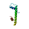
| ||||||||
|---|---|---|---|---|---|---|---|---|---|
| 1 | 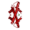
| ||||||||
| Unit cell |
|
-Components
| #1: Protein | Mass: 7683.054 Da / Num. of mol.: 1 Source method: isolated from a genetically manipulated source Source: (gene. exp.) Methanothermus fervidus (archaea) / Gene: HMFB / Gene (production host): HMFB / Production host:  |
|---|---|
| #2: Chemical | ChemComp-ZN /   Mass: 65.409 Da / Num. of mol.: 1 / Source method: obtained synthetically / Formula: Zn Mass: 65.409 Da / Num. of mol.: 1 / Source method: obtained synthetically / Formula: Zn |
| #3: Chemical | ChemComp-CL /   Mass: 35.453 Da / Num. of mol.: 1 / Source method: obtained synthetically / Formula: Cl Mass: 35.453 Da / Num. of mol.: 1 / Source method: obtained synthetically / Formula: Cl |
| #4: Water | ChemComp-HOH /  Mass: 18.015 Da / Num. of mol.: 46 / Source method: isolated from a natural source / Formula: H2O Mass: 18.015 Da / Num. of mol.: 46 / Source method: isolated from a natural source / Formula: H2O |
-Experimental details
-Experiment
| Experiment | Method: X-RAY DIFFRACTION / Number of used crystals: 1 |
|---|
- Sample preparation
| Crystal | Density Matthews: 2.5 Å3/Da / Density % sol: 51 % Description: ORTHOROMBIC HMFA STRUCTURE WILL BE SUBMITTED SOON | ||||||||||||||||||||
|---|---|---|---|---|---|---|---|---|---|---|---|---|---|---|---|---|---|---|---|---|---|
| Crystal grow | *PLUS pH: 8 / Method: vapor diffusion, hanging drop | ||||||||||||||||||||
| Components of the solutions | *PLUS
|
-Data collection
| Diffraction | Mean temperature: 293 K |
|---|---|
| Diffraction source | Source: ROTATING ANODE / Type: RIGAKU RUH2R / Wavelength: 1.5418 |
| Detector | Type: MAR scanner 300 mm plate / Detector: IMAGE PLATE / Date: Feb 1, 1995 / Details: SUPPER NI-COATED MIRROR SYSTEM |
| Radiation | Monochromator: NI FOIL / Monochromatic (M) / Laue (L): M / Scattering type: x-ray |
| Radiation wavelength | Wavelength: 1.5418 Å / Relative weight: 1 |
| Reflection | Resolution: 1.9→39.1 Å / Num. obs: 6152 / % possible obs: 95.9 % / Observed criterion σ(I): 0 / Redundancy: 4.1 % / Biso Wilson estimate: 22.87 Å2 / Rmerge(I) obs: 0.053 / Rsym value: 0.053 / Net I/σ(I): 8.3 |
| Reflection shell | Resolution: 1.9→1.97 Å / Redundancy: 3.4 % / Rmerge(I) obs: 0.196 / Mean I/σ(I) obs: 3.7 / Rsym value: 0.196 / % possible all: 97.2 |
| Reflection | *PLUS Num. measured all: 25200 |
| Reflection shell | *PLUS % possible obs: 97.2 % |
- Processing
| Software |
| ||||||||||||||||||||||||||||||||||||||||||||||||||||||||||||||||||||||||||||||||||||
|---|---|---|---|---|---|---|---|---|---|---|---|---|---|---|---|---|---|---|---|---|---|---|---|---|---|---|---|---|---|---|---|---|---|---|---|---|---|---|---|---|---|---|---|---|---|---|---|---|---|---|---|---|---|---|---|---|---|---|---|---|---|---|---|---|---|---|---|---|---|---|---|---|---|---|---|---|---|---|---|---|---|---|---|---|---|
| Refinement | Method to determine structure: MOLECULAR REPLACEMENT Starting model: PARTIALLY REFINED ORTHORHOMBIC HMFA Resolution: 1.55→18 Å / Cross valid method: AFTER RIGID BODY REFINEMENT / σ(F): 0 Details: RIGID BODY WITH AMORE, SIMULATED ANNEALING WITH X-PLOR, REFINEMENT WITH REFMAC. ESD FROM LUZZATI PLOT (A) : 0.25
| ||||||||||||||||||||||||||||||||||||||||||||||||||||||||||||||||||||||||||||||||||||
| Displacement parameters | Biso mean: 25.34 Å2
| ||||||||||||||||||||||||||||||||||||||||||||||||||||||||||||||||||||||||||||||||||||
| Refine analyze | Luzzati coordinate error obs: 0.25 Å | ||||||||||||||||||||||||||||||||||||||||||||||||||||||||||||||||||||||||||||||||||||
| Refinement step | Cycle: LAST / Resolution: 1.55→18 Å
| ||||||||||||||||||||||||||||||||||||||||||||||||||||||||||||||||||||||||||||||||||||
| Refine LS restraints |
| ||||||||||||||||||||||||||||||||||||||||||||||||||||||||||||||||||||||||||||||||||||
| Software | *PLUS Name: REFMAC / Classification: refinement | ||||||||||||||||||||||||||||||||||||||||||||||||||||||||||||||||||||||||||||||||||||
| Refinement | *PLUS Highest resolution: 1.9 Å / Num. reflection obs: 5835 / Num. reflection Rfree: 290 / Rfactor obs: 0.18 / Rfactor Rfree: 0.229 | ||||||||||||||||||||||||||||||||||||||||||||||||||||||||||||||||||||||||||||||||||||
| Solvent computation | *PLUS | ||||||||||||||||||||||||||||||||||||||||||||||||||||||||||||||||||||||||||||||||||||
| Displacement parameters | *PLUS | ||||||||||||||||||||||||||||||||||||||||||||||||||||||||||||||||||||||||||||||||||||
| Refine LS restraints | *PLUS
|
