 Movie
Movie Controller

[English] 日本語
 Yorodumi
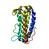
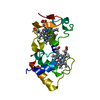
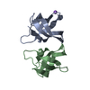
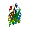
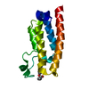
- PDB-1a62: CRYSTAL STRUCTURE OF THE RNA-BINDING DOMAIN OF THE TRANSCRIPTIONA... -
+ Open data

- Basic information
| Entry | Database: PDB / ID: 1a62 | ||||||
|---|---|---|---|---|---|---|---|
| Title | CRYSTAL STRUCTURE OF THE RNA-BINDING DOMAIN OF THE TRANSCRIPTIONAL TERMINATOR PROTEIN RHO | ||||||
 Components Components | RHO | ||||||
 Keywords Keywords | TRANSCRIPTION TERMINATION / TERMINATION / RNA BINDING DOMAIN / TRANSCRIPTION REGULATION / OB FOLD / F1-ATPASE | ||||||
| Function / homology |  Function and homology information Function and homology informationATP-dependent activity, acting on RNA / DNA-templated transcription termination / helicase activity / Hydrolases; Acting on acid anhydrides; Acting on acid anhydrides to facilitate cellular and subcellular movement / hydrolase activity / ATP hydrolysis activity / RNA binding / ATP binding / identical protein binding / membrane / cytosol Similarity search - Function | ||||||
| Biological species |  | ||||||
| Method |  X-RAY DIFFRACTION / SYNCHROTRON / MAD / Resolution: 1.55 Å X-RAY DIFFRACTION / SYNCHROTRON / MAD / Resolution: 1.55 Å | ||||||
 Authors Authors | Allison, T.J. / Wood, T.C. / Briercheck, D.M. / Rastinejad, F. / Richardson, J.P. / Rule, G.S. | ||||||
 Citation Citation | Journal: Nat.Struct.Biol. / Year: 1998 Title: Crystal structure of the RNA-binding domain from transcription termination factor rho. Authors: Allison, T.J. / Wood, T.C. / Briercheck, D.M. / Rastinejad, F. / Richardson, J.P. / Rule, G.S. #1: Journal: Nat.Struct.Biol. / Year: 1998Title: The NMR Structure of the RNA Binding Domain of E. Coli Rho Factor Suggests Possible RNA-Protein Interactions Authors: Briercheck, D.M. / Wood, T.C. / Allison, T.J. / Richardson, J.P. / Rule, G.S. #2: Journal: J.Biomol.NMR / Year: 1996Title: 1H, 15N and 13C Resonance Assignments and Secondary Structure Determination of the RNA-Binding Domain of E.Coli Rho Protein Authors: Briercheck, D.M. / Allison, T.J. / Richardson, J.P. / Ellena, J.F. / Wood, T.C. / Rule, G.S. | ||||||
| History |
|
- Structure visualization
| Structure viewer | Molecule: MolmilJmol/JSmol |
|---|
- Downloads & links
-Download
| PDBx/mmCIF format | 1a62.cif.gz | 39.9 KB | Display | PDBx/mmCIF format |
|---|---|---|---|---|
| PDB format | pdb1a62.ent.gz | 27 KB | Display | PDB format |
| PDBx/mmJSON format | 1a62.json.gz | Tree view | PDBx/mmJSON format | |
| Others |  Other downloads Other downloads |
-Validation report
| Summary document | 1a62_validation.pdf.gz | 362.2 KB | Display | wwPDB validaton report |
|---|---|---|---|---|
| Full document | 1a62_full_validation.pdf.gz | 364 KB | Display | |
| Data in XML | 1a62_validation.xml.gz | 4 KB | Display | |
| Data in CIF | 1a62_validation.cif.gz | 6.2 KB | Display | |
| Arichive directory | https://data.pdbj.org/pub/pdb/validation_reports/a6/1a62ftp://data.pdbj.org/pub/pdb/validation_reports/a6/1a62 | HTTPS FTP |
-Related structure data
| Similar structure data |
|---|


-Links
 PDBj
PDBj


- Assembly
| Deposited unit | 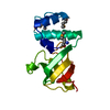
| ||||||||
|---|---|---|---|---|---|---|---|---|---|
| 1 |
| ||||||||
| Unit cell |
|
-Components
| #1: Protein | Mass: 14775.287 Da / Num. of mol.: 1 / Fragment: RNA BINDING DOMAIN, RESIDUES 1 - 130 Source method: isolated from a genetically manipulated source Source: (gene. exp.) |
|---|---|
| #2: Water | ChemComp-HOH /  Mass: 18.015 Da / Num. of mol.: 115 / Source method: isolated from a natural source / Formula: H2O Mass: 18.015 Da / Num. of mol.: 115 / Source method: isolated from a natural source / Formula: H2O |
| Has protein modification | Y |
-Experimental details
-Experiment
| Experiment | Method: X-RAY DIFFRACTION / Number of used crystals: 4 |
|---|
- Sample preparation
| Crystal | Density Matthews: 2.08 Å3/Da / Density % sol: 33 % | |||||||||||||||
|---|---|---|---|---|---|---|---|---|---|---|---|---|---|---|---|---|
| Crystal grow | pH: 7 / Details: pH 7.0 | |||||||||||||||
| Crystal grow | *PLUS Method: microdialysis | |||||||||||||||
| Components of the solutions | *PLUS
|
-Data collection
| Diffraction | Mean temperature: 100 K | |||||||||||||||
|---|---|---|---|---|---|---|---|---|---|---|---|---|---|---|---|---|
| Diffraction source | Source: SYNCHROTRON / Site: NSLS  / Beamline: X4A / Wavelength: 0.969, 0.978, 0.979, 0.984 / Beamline: X4A / Wavelength: 0.969, 0.978, 0.979, 0.984 | |||||||||||||||
| Detector | Type: FUJI / Detector: IMAGE PLATE / Date: May 1, 1997 / Details: MIRROR | |||||||||||||||
| Radiation | Monochromator: SI(111) / Monochromatic (M) / Laue (L): M / Scattering type: x-ray | |||||||||||||||
| Radiation wavelength |
| |||||||||||||||
| Reflection | Resolution: 1.55→30 Å / Num. obs: 16360 / % possible obs: 92 % / Observed criterion σ(I): 0.01 / Redundancy: 6 % / Rmerge(I) obs: 0.068 / Rsym value: 0.04 / Net I/σ(I): 22 | |||||||||||||||
| Reflection shell | Resolution: 1.55→1.61 Å / Redundancy: 5 % / Rmerge(I) obs: 0.27 / Mean I/σ(I) obs: 5.2 / Rsym value: 0.26 / % possible all: 67 | |||||||||||||||
| Reflection shell | *PLUS % possible obs: 67 % |
- Processing
| Software |
| ||||||||||||||||||||||||||||||||||||||||||||||||||||||||||||
|---|---|---|---|---|---|---|---|---|---|---|---|---|---|---|---|---|---|---|---|---|---|---|---|---|---|---|---|---|---|---|---|---|---|---|---|---|---|---|---|---|---|---|---|---|---|---|---|---|---|---|---|---|---|---|---|---|---|---|---|---|---|
| Refinement | Method to determine structure: MAD / Resolution: 1.55→30 Å / Isotropic thermal model: RESTRAINED / Cross valid method: THROUGHOUT / σ(F): 0.01
| ||||||||||||||||||||||||||||||||||||||||||||||||||||||||||||
| Displacement parameters | Biso mean: 21.1 Å2 | ||||||||||||||||||||||||||||||||||||||||||||||||||||||||||||
| Refinement step | Cycle: LAST / Resolution: 1.55→30 Å
| ||||||||||||||||||||||||||||||||||||||||||||||||||||||||||||
| Refine LS restraints |
| ||||||||||||||||||||||||||||||||||||||||||||||||||||||||||||
| Xplor file |
| ||||||||||||||||||||||||||||||||||||||||||||||||||||||||||||
| Software | *PLUS Name: X-PLOR / Version: 3.851 / Classification: refinement | ||||||||||||||||||||||||||||||||||||||||||||||||||||||||||||
| Refinement | *PLUS | ||||||||||||||||||||||||||||||||||||||||||||||||||||||||||||
| Solvent computation | *PLUS | ||||||||||||||||||||||||||||||||||||||||||||||||||||||||||||
| Displacement parameters | *PLUS |
