 Movie
Movie Controller


+ Open data

- Basic information
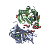
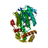
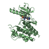
| Entry | Database: PDB / ID: 1a3a | ||||||
|---|---|---|---|---|---|---|---|
| Title | CRYSTAL STRUCTURE OF IIA MANNITOL FROM ESCHERICHIA COLI | ||||||
 Components Components | MANNITOL-SPECIFIC EII | ||||||
 Keywords Keywords | PHOSPHOTRANSFERASE / PHOSPHOENOLPYRUVATE DEPENDENT PHOSPHOTRANSFERASE SYSTEM / IIA ENZYMES / HISTIDINE PHOSPHORYLATION | ||||||
| Function / homology |  Function and homology information Function and homology informationprotein-Npi-phosphohistidine-D-mannitol phosphotransferase / protein-N(PI)-phosphohistidine-mannitol phosphotransferase system transmembrane transporter activity / protein-phosphocysteine-mannitol phosphotransferase system transporter activity / sorbitol transmembrane transport / mannitol transmembrane transport / protein-phosphocysteine-sugar phosphotransferase activity / phosphoenolpyruvate-dependent sugar phosphotransferase system / kinase activity / plasma membrane Similarity search - Function | ||||||
| Biological species |  | ||||||
| Method |  X-RAY DIFFRACTION / SYNCHROTRON / MULTIPLE ANOMALOUS DISPERSION / Resolution: 1.8 Å X-RAY DIFFRACTION / SYNCHROTRON / MULTIPLE ANOMALOUS DISPERSION / Resolution: 1.8 Å | ||||||
 Authors Authors | Van Montfort, R.L.M. / Pijning, T. / Kalk, K.H. / Hangyi, I. / Kouwijzer, M.L.C.E. / Robillard, G.T. / Dijkstra, B.W. | ||||||
 Citation Citation | Journal: Structure / Year: 1998 Title: The structure of the Escherichia coli phosphotransferase IIAmannitol reveals a novel fold with two conformations of the active site. Authors: van Montfort, R.L. / Pijning, T. / Kalk, K.H. / Hangyi, I. / Kouwijzer, M.L. / Robillard, G.T. / Dijkstra, B.W. | ||||||
| History |
|
- Structure visualization
| Structure viewer | Molecule: MolmilJmol/JSmol |
|---|
- Downloads & links
-Download
| PDBx/mmCIF format | 1a3a.cif.gz | 130.1 KB | Display | PDBx/mmCIF format |
|---|---|---|---|---|
| PDB format | pdb1a3a.ent.gz | 102.6 KB | Display | PDB format |
| PDBx/mmJSON format | 1a3a.json.gz | Tree view | PDBx/mmJSON format | |
| Others |  Other downloads Other downloads |
-Validation report
| Arichive directory | https://data.pdbj.org/pub/pdb/validation_reports/a3/1a3aftp://data.pdbj.org/pub/pdb/validation_reports/a3/1a3a | HTTPS FTP |
|---|
-Related structure data
| Similar structure data |
|---|


-Links
 PDBj
PDBj- Assembly
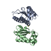
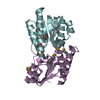
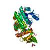
| Deposited unit | 
| ||||||||||||||||
|---|---|---|---|---|---|---|---|---|---|---|---|---|---|---|---|---|---|
| 1 | 
| ||||||||||||||||
| 2 | 
| ||||||||||||||||
| Unit cell |
| ||||||||||||||||
| Noncrystallographic symmetry (NCS) | NCS oper:
|
-Components
| #1: Protein | Mass: 16348.547 Da / Num. of mol.: 4 / Fragment: IIA DOMAIN, RESIDUES 491 - 637 Source method: isolated from a genetically manipulated source Source: (gene. exp.) References: UniProt: P00550, protein-Npi-phosphohistidine-sugar phosphotransferase #2: Water | ChemComp-HOH / |  Mass: 18.015 Da / Num. of mol.: 568 / Source method: isolated from a natural source / Formula: H2O Mass: 18.015 Da / Num. of mol.: 568 / Source method: isolated from a natural source / Formula: H2O |
|---|
-Experimental details
-Experiment
| Experiment | Method: X-RAY DIFFRACTION / Number of used crystals: 1 |
|---|
- Sample preparation
| Crystal | Density Matthews: 1.9 Å3/Da / Density % sol: 36.4 % Description: MAD DATA WERE COLLECTED AT THE BM14 AT THE ESRF, GRENOBLE | ||||||||||||||||||||||||||||||||||||||||||||||||||||||||||||
|---|---|---|---|---|---|---|---|---|---|---|---|---|---|---|---|---|---|---|---|---|---|---|---|---|---|---|---|---|---|---|---|---|---|---|---|---|---|---|---|---|---|---|---|---|---|---|---|---|---|---|---|---|---|---|---|---|---|---|---|---|---|
| Crystal grow | pH: 7.5 / Details: pH 7.5 | ||||||||||||||||||||||||||||||||||||||||||||||||||||||||||||
| Crystal | *PLUS | ||||||||||||||||||||||||||||||||||||||||||||||||||||||||||||
| Crystal grow | *PLUS Temperature: 5 ℃ / Method: vapor diffusion | ||||||||||||||||||||||||||||||||||||||||||||||||||||||||||||
| Components of the solutions | *PLUS
|
-Data collection
| Diffraction | Mean temperature: 120 K |
|---|---|
| Diffraction source | Source: SYNCHROTRON / Site: EMBL/DESY, HAMBURG  / Beamline: X11 / Wavelength: 0.92 / Beamline: X11 / Wavelength: 0.92 |
| Detector | Type: MARRESEARCH / Detector: IMAGE PLATE / Date: Feb 1, 1997 |
| Radiation | Monochromatic (M) / Laue (L): M / Scattering type: x-ray |
| Radiation wavelength | Wavelength: 0.92 Å / Relative weight: 1 |
| Reflection | Highest resolution: 1.8 Å / Num. obs: 45271 / % possible obs: 95.8 % / Observed criterion σ(I): 2 / Redundancy: 4.3 % / Rmerge(I) obs: 0.055 / Net I/σ(I): 14.4 |
| Reflection shell | Resolution: 1.8→1.83 Å / Rmerge(I) obs: 0.262 / % possible all: 84.2 |
| Reflection | *PLUS Num. measured all: 195844 |
| Reflection shell | *PLUS % possible obs: 84.2 % |
- Processing
| Software |
| ||||||||||||||||
|---|---|---|---|---|---|---|---|---|---|---|---|---|---|---|---|---|---|
| Refinement | Method to determine structure: MULTIPLE ANOMALOUS DISPERSION Resolution: 1.8→5 Å / Cross valid method: THROUGHOUT
| ||||||||||||||||
| Refinement step | Cycle: LAST / Resolution: 1.8→5 Å
| ||||||||||||||||
| Software | *PLUS Name: X-PLOR / Classification: refinement | ||||||||||||||||
| Refinement | *PLUS Rfactor all: 0.185 / Rfactor obs: 0.19 / Rfactor Rwork: 0.19 | ||||||||||||||||
| Solvent computation | *PLUS | ||||||||||||||||
| Displacement parameters | *PLUS | ||||||||||||||||
| Refine LS restraints | *PLUS
|
