 Movie
Movie Controller


+ Open data

- Basic information
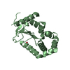
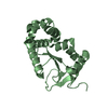
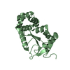
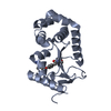
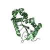
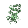
| Entry | Database: PDB / ID: 1a2m | ||||||
|---|---|---|---|---|---|---|---|
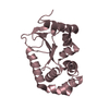
| Title | OXIDIZED DSBA AT 2.7 ANGSTROMS RESOLUTION, CRYSTAL FORM III | ||||||
 Components Components | DISULFIDE BOND FORMATION PROTEIN | ||||||
 Keywords Keywords | OXIDOREDUCTASE / PROTEIN DISULFIDE ISOMERASE / PROTEIN FOLDING / REDOX PROTEIN / REDOX-ACTIVE CENTER | ||||||
| Function / homology |  Function and homology information Function and homology informationcellular response to antibiotic / protein disulfide isomerase activity / protein-disulfide reductase activity / outer membrane-bounded periplasmic space / oxidoreductase activity / periplasmic space Similarity search - Function | ||||||
| Biological species |  | ||||||
| Method |  X-RAY DIFFRACTION / MOLECULAR REPLACEMENT / Resolution: 2.7 Å X-RAY DIFFRACTION / MOLECULAR REPLACEMENT / Resolution: 2.7 Å | ||||||
 Authors Authors | Martin, J.L. / Guddat, L.W. | ||||||
 Citation Citation | Journal: Structure / Year: 1998 Title: Crystal structures of reduced and oxidized DsbA: investigation of domain motion and thiolate stabilization. Authors: Guddat, L.W. / Bardwell, J.C. / Martin, J.L. #1: Journal: Protein Sci. / Year: 1997Title: Structural Analysis of Three His32 Mutants of Dsba: Support for an Electrostatic Role of His32 in Dsba Stability Authors: Guddat, L.W. / Bardwell, J.C. / Glockshuber, R. / Huber-Wunderlich, M. / Zander, T. / Martin, J.L. #2: Journal: Protein Sci. / Year: 1997Title: The Uncharged Surface Features Surrounding the Active Site of Escherichia Coli Dsba are Conserved and are Implicated in Peptide Binding Authors: Guddat, L.W. / Bardwell, J.C. / Zander, T. / Martin, J.L. #3: Journal: Nature / Year: 1993Title: Crystal Structure of the Dsba Protein Required for Disulphide Bond Formation in Vivo Authors: Martin, J.L. / Bardwell, J.C. / Kuriyan, J. #4: Journal: J.Mol.Biol. / Year: 1993Title: Crystallization of Dsba, an Escherichia Coli Protein Required for Disulphide Bond Formation in Vivo Authors: Martin, J.L. / Waksman, G. / Bardwell, J.C. / Beckwith, J. / Kuriyan, J. | ||||||
| History |
|
- Structure visualization
| Structure viewer | Molecule: MolmilJmol/JSmol |
|---|
- Downloads & links
-Download
| PDBx/mmCIF format | 1a2m.cif.gz | 81.2 KB | Display | PDBx/mmCIF format |
|---|---|---|---|---|
| PDB format | pdb1a2m.ent.gz | 61.2 KB | Display | PDB format |
| PDBx/mmJSON format | 1a2m.json.gz | Tree view | PDBx/mmJSON format | |
| Others |  Other downloads Other downloads |
-Validation report
| Summary document | 1a2m_validation.pdf.gz | 433.4 KB | Display | wwPDB validaton report |
|---|---|---|---|---|
| Full document | 1a2m_full_validation.pdf.gz | 437.3 KB | Display | |
| Data in XML | 1a2m_validation.xml.gz | 15.1 KB | Display | |
| Data in CIF | 1a2m_validation.cif.gz | 19.9 KB | Display | |
| Arichive directory | https://data.pdbj.org/pub/pdb/validation_reports/a2/1a2mftp://data.pdbj.org/pub/pdb/validation_reports/a2/1a2m | HTTPS FTP |
-Related structure data
| Related structure data | 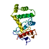 1a2jC 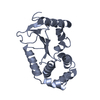 1a2lC  1fvkS S: Starting model for refinement C: citing same article ( |
|---|---|
| Similar structure data |


-Links
 PDBj
PDBj

- Assembly
| Deposited unit | 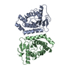
| ||||||||
|---|---|---|---|---|---|---|---|---|---|
| 1 | 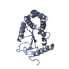
| ||||||||
| 2 | 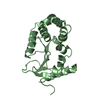
| ||||||||
| Unit cell |
|
-Components
| #1: Protein | Mass: 21155.025 Da / Num. of mol.: 2 Source method: isolated from a genetically manipulated source Source: (gene. exp.) #2: Water | ChemComp-HOH / |  Mass: 18.015 Da / Num. of mol.: 25 / Source method: isolated from a natural source / Formula: H2O Mass: 18.015 Da / Num. of mol.: 25 / Source method: isolated from a natural source / Formula: H2OHas protein modification | Y | |
|---|
-Experimental details
-Experiment
| Experiment | Method: X-RAY DIFFRACTION / Number of used crystals: 1 |
|---|
- Sample preparation
| Crystal | Density Matthews: 2.61 Å3/Da / Density % sol: 49 % | ||||||||||||||||||||||||||||||||||||||||
|---|---|---|---|---|---|---|---|---|---|---|---|---|---|---|---|---|---|---|---|---|---|---|---|---|---|---|---|---|---|---|---|---|---|---|---|---|---|---|---|---|---|
| Crystal grow | pH: 5.6 Details: 0.2 M AMMONIUM ACETATE, 0.1M SODIUM CITRATE PH 5.6 30% (W/V) PEG 4000. | ||||||||||||||||||||||||||||||||||||||||
| Crystal | *PLUS | ||||||||||||||||||||||||||||||||||||||||
| Crystal grow | *PLUS Temperature: 21 ℃ / pH: 6.5 / Method: vapor diffusion, hanging drop / Details: Martin, J.L., (1993) J.Mol.Biol., 230, 1097. | ||||||||||||||||||||||||||||||||||||||||
| Components of the solutions | *PLUS
|
-Data collection
| Diffraction | Mean temperature: 289 K |
|---|---|
| Diffraction source | Source: ROTATING ANODE / Type: RIGAKU RUH2R / Wavelength: 1.5418 |
| Detector | Type: RIGAKU RAXIS IIC / Detector: IMAGE PLATE / Date: Dec 10, 1996 / Details: YALE MIRRORS |
| Radiation | Monochromator: GRAPHITE(002) / Monochromatic (M) / Laue (L): M / Scattering type: x-ray |
| Radiation wavelength | Wavelength: 1.5418 Å / Relative weight: 1 |
| Reflection | Resolution: 2.7→50 Å / Num. obs: 18501 / % possible obs: 73.6 % / Observed criterion σ(I): 1 / Redundancy: 2 % / Rmerge(I) obs: 0.118 / Rsym value: 0.118 / Net I/σ(I): 7 |
| Reflection shell | Resolution: 2.7→2.8 Å / Redundancy: 1.4 % / Rmerge(I) obs: 0.294 / Mean I/σ(I) obs: 2.2 / Rsym value: 0.294 / % possible all: 56.1 |
| Reflection | *PLUS Num. obs: 10864 / % possible obs: 86.1 % / Num. measured all: 43081 / Rmerge(I) obs: 0.083 |
| Reflection shell | *PLUS % possible obs: 69.9 % / Rmerge(I) obs: 0.298 / Mean I/σ(I) obs: 3.2 |
- Processing
| Software |
| ||||||||||||||||||||||||||||||||||||||||||||||||||||||||||||||||||||||||||||||||
|---|---|---|---|---|---|---|---|---|---|---|---|---|---|---|---|---|---|---|---|---|---|---|---|---|---|---|---|---|---|---|---|---|---|---|---|---|---|---|---|---|---|---|---|---|---|---|---|---|---|---|---|---|---|---|---|---|---|---|---|---|---|---|---|---|---|---|---|---|---|---|---|---|---|---|---|---|---|---|---|---|---|
| Refinement | Method to determine structure: MOLECULAR REPLACEMENT Starting model: PDB ENTRY 1FVK Resolution: 2.7→50 Å / Data cutoff high absF: 1000000 / Data cutoff low absF: 0.001 / σ(F): 1
| ||||||||||||||||||||||||||||||||||||||||||||||||||||||||||||||||||||||||||||||||
| Displacement parameters | Biso mean: 24.2 Å2 | ||||||||||||||||||||||||||||||||||||||||||||||||||||||||||||||||||||||||||||||||
| Refine analyze |
| ||||||||||||||||||||||||||||||||||||||||||||||||||||||||||||||||||||||||||||||||
| Refinement step | Cycle: LAST / Resolution: 2.7→50 Å
| ||||||||||||||||||||||||||||||||||||||||||||||||||||||||||||||||||||||||||||||||
| Refine LS restraints |
| ||||||||||||||||||||||||||||||||||||||||||||||||||||||||||||||||||||||||||||||||
| LS refinement shell | Resolution: 2.7→2.8 Å / Total num. of bins used: 8
| ||||||||||||||||||||||||||||||||||||||||||||||||||||||||||||||||||||||||||||||||
| Xplor file | Serial no: 1 / Param file: PARHCSDX.PRO / Topol file: TOPHCSDX.PRO | ||||||||||||||||||||||||||||||||||||||||||||||||||||||||||||||||||||||||||||||||
| Software | *PLUS Name: X-PLOR / Version: 3.851 / Classification: refinement | ||||||||||||||||||||||||||||||||||||||||||||||||||||||||||||||||||||||||||||||||
| Refine LS restraints | *PLUS
|
