[English] 日本語
Yorodumi
- PDB-1a1q: HEPATITIS C VIRUS NS3 PROTEINASE -

+
Open data


ID or keywords:

Loading...

-
Basic information

Entry
Database: PDB / ID: 1a1q
TitleHEPATITIS C VIRUS NS3 PROTEINASE
ComponentsNS3 PROTEINASE
KeywordsHYDROLASE / SERINE PROTEASE
Function / homology
Function and homology information


hepacivirin / host cell mitochondrial membrane / host cell lipid droplet / symbiont-mediated suppression of host TRAF-mediated signal transduction / transformation of host cell by virus / symbiont-mediated perturbation of host cell cycle G1/S transition checkpoint / symbiont-mediated suppression of host JAK-STAT cascade via inhibition of STAT1 activity / symbiont-mediated suppression of host cytoplasmic pattern recognition receptor signaling pathway via inhibition of MAVS activity / SH3 domain binding / : ...hepacivirin / host cell mitochondrial membrane / host cell lipid droplet / symbiont-mediated suppression of host TRAF-mediated signal transduction / transformation of host cell by virus / symbiont-mediated perturbation of host cell cycle G1/S transition checkpoint / symbiont-mediated suppression of host JAK-STAT cascade via inhibition of STAT1 activity / symbiont-mediated suppression of host cytoplasmic pattern recognition receptor signaling pathway via inhibition of MAVS activity / SH3 domain binding / : / nucleoside-triphosphate phosphatase / protein complex oligomerization / monoatomic ion channel activity / clathrin-dependent endocytosis of virus by host cell / viral nucleocapsid / Hydrolases; Acting on peptide bonds (peptidases); Cysteine endopeptidases / RNA helicase activity / host cell endoplasmic reticulum membrane / host cell perinuclear region of cytoplasm / RNA helicase / induction by virus of host autophagy / RNA-directed RNA polymerase / ribonucleoprotein complex / viral RNA genome replication / cysteine-type endopeptidase activity / RNA-dependent RNA polymerase activity / serine-type endopeptidase activity / fusion of virus membrane with host endosome membrane / viral envelope / symbiont-mediated suppression of host type I interferon-mediated signaling pathway / host cell nucleus / structural molecule activity / virion attachment to host cell / host cell plasma membrane / virion membrane / ATP hydrolysis activity / proteolysis / RNA binding / zinc ion binding / ATP binding / membrane
Similarity search - Function
Hepatitis C virus core protein, chain A superfamily / Hepatitus C virus, Non-structural 5a protein, C-terminal / Hepatitis C virus NS5A, 1B domain superfamily / Hepatitis C virus non-structural protein NS2, C-terminal domain / Hepatitis C virus non-structural protein NS2, N-terminal domain / Hepatitis C virus non-structural protein NS2 / HCV NS5a protein C-terminal region / Hepatitis C virus, Non-structural protein NS4b / Hepatitis C virus, Core protein, N-terminal / Hepatitis C virus non-structural protein NS4b ...Hepatitis C virus core protein, chain A superfamily / Hepatitus C virus, Non-structural 5a protein, C-terminal / Hepatitis C virus NS5A, 1B domain superfamily / Hepatitis C virus non-structural protein NS2, C-terminal domain / Hepatitis C virus non-structural protein NS2, N-terminal domain / Hepatitis C virus non-structural protein NS2 / HCV NS5a protein C-terminal region / Hepatitis C virus, Non-structural protein NS4b / Hepatitis C virus, Core protein, N-terminal / Hepatitis C virus non-structural protein NS4b / Hepatitis C virus capsid protein / Hepatitis C virus, Non-structural protein NS2 / Hepatitis C virus, Non-structural 5a protein / Hepatitis C virus, Non-structural 5a protein, domain 1a / Hepatitis C virus non-structural 5a, 1B domain / NS5A domain 1a superfamily / Hepatitis C virus non-structural 5a protein membrane anchor / Hepatitis C virus non-structural 5a zinc finger domain / Hepatitis C virus non-structural 5a domain 1b / Hepacivirus nonstructural protein 2 (NS2) protease domain profile. / Hepatitis C virus, Non-structural protein NS4a / Hepatitis C virus non-structural protein NS4a / Hepatitis C virus, Core protein, C-terminal / Hepatitis C virus core protein / Hepatitis C virus, Non-structural protein E2/NS1 / Hepatitis C virus non-structural protein E2/NS1 / Hepatitis C virus, Envelope glycoprotein E1 / Hepatitis C virus envelope glycoprotein E1 / RNA dependent RNA polymerase, hepatitis C virus / Viral RNA dependent RNA polymerase / Hepatitis C virus, NS3 protease, Peptidase S29 / Hepatitis C virus NS3 protease / Hepacivirus/Pegivirus NS3 protease domain profile. / DEAD box, Flavivirus / Flavivirus DEAD domain / Superfamilies 1 and 2 helicase C-terminal domain profile. / Superfamilies 1 and 2 helicase ATP-binding type-1 domain profile. / DEAD-like helicases superfamily / Helicase, C-terminal / Helicase superfamily 1/2, ATP-binding domain / Reverse transcriptase/Diguanylate cyclase domain / RNA-directed RNA polymerase, catalytic domain / RdRp of positive ssRNA viruses catalytic domain profile. / Peptidase S1, PA clan, chymotrypsin-like fold / Peptidase S1, PA clan / DNA/RNA polymerase superfamily / P-loop containing nucleoside triphosphate hydrolase
Similarity search - Domain/homology
Biological speciesHepatitis C virus
MethodX-RAY DIFFRACTION / SYNCHROTRON / HEAVY ATOMS: ISOMORPHOUS, ANOMALOUS SIGNALS / Resolution: 2.4 Å
AuthorsLove, R.A. / Parge, H.E. / Wickersham, J.A. / Hostomsky, Z. / Habuka, N. / Moomaw, E.W. / Adachi, T. / Hostomska, Z.
CitationJournal: Cell(Cambridge,Mass.) / Year: 1996
Title: The crystal structure of hepatitis C virus NS3 proteinase reveals a trypsin-like fold and a structural zinc binding site.
Authors: Love, R.A. / Parge, H.E. / Wickersham, J.A. / Hostomsky, Z. / Habuka, N. / Moomaw, E.W. / Adachi, T. / Hostomska, Z.
History
DepositionDec 12, 1997Processing site: BNL
Revision 1.0Mar 25, 1998Provider: repository / Type: Initial release
Revision 1.1Mar 24, 2008Group: Version format compliance
Revision 1.2Jul 13, 2011Group: Version format compliance
Revision 1.3Feb 7, 2024Group: Data collection / Database references / Derived calculations
Category: chem_comp_atom / chem_comp_bond ...chem_comp_atom / chem_comp_bond / database_2 / struct_ref_seq_dif / struct_site
Item: _database_2.pdbx_DOI / _database_2.pdbx_database_accession ..._database_2.pdbx_DOI / _database_2.pdbx_database_accession / _struct_ref_seq_dif.details / _struct_site.pdbx_auth_asym_id / _struct_site.pdbx_auth_comp_id / _struct_site.pdbx_auth_seq_id

-
Structure visualization

Structure viewerMolecule:
MolmilJmol/JSmol

Downloads & links

-
Assembly

Deposited unit
A: NS3 PROTEINASE
B: NS3 PROTEINASE
C: NS3 PROTEINASE
hetero molecules


Theoretical massNumber of molelcules
Total (without water)59,8806
Polymers59,6843
Non-polymers1963
Water0
1
A: NS3 PROTEINASE
hetero molecules


Theoretical massNumber of molelcules
Total (without water)19,9602
Polymers19,8951
Non-polymers651
Water0
TypeNameSymmetry operationNumber
identity operation1_555x,y,z1
2
B: NS3 PROTEINASE
hetero molecules


Theoretical massNumber of molelcules
Total (without water)19,9602
Polymers19,8951
Non-polymers651
Water0
TypeNameSymmetry operationNumber
identity operation1_555x,y,z1
3
C: NS3 PROTEINASE
hetero molecules


Theoretical massNumber of molelcules
Total (without water)19,9602
Polymers19,8951
Non-polymers651
Water0
TypeNameSymmetry operationNumber
identity operation1_555x,y,z1
Unit cell
Length a, b, c (Å)133.000, 133.000, 223.000
Angle α, β, γ (deg.)90.00, 90.00, 120.00
Int Tables number155
Space group name H-MH32
Noncrystallographic symmetry (NCS)NCS oper:
IDCodeMatrixVector
1given(-0.95206, 0.305556, -0.014748), (-0.305663, -0.952124, 0.005574), (-0.012339, 0.009815, 0.999876)-22.67001, 73.03197, 0.78307
2given(-0.919307, -0.392193, 0.032539), (-0.388774, 0.917892, 0.079553), (-0.061068, 0.060484, -0.9963)27.04836, 2.94648, -2.43504

-
Components

#1: Protein NS3 PROTEINASE / Coordinate model: Cα atoms only


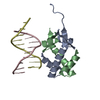
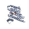
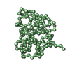
Mass: 19894.604 Da / Num. of mol.: 3
Source method: isolated from a genetically manipulated source
Source: (gene. exp.) Hepatitis C virus / Genus: Hepacivirus / Strain: TYPE 1B / Description: EXPRESSED AS SOLUBLE PROTEIN / Gene: CDNA / Variant: BK ISOLATE / Plasmid: CDNA DERIVED FROM VIRAL RNA ISOLATED FROM PAT / Gene (production host): CDNA DERIVED FROM / Production host: Escherichia coli (E. coli) / References: UniProt: P26663
#2: Chemical ChemComp-ZN / ZINC ION


Mass: 65.409 Da / Num. of mol.: 3 / Source method: obtained synthetically / Formula: Zn

-
Experimental details

-
Experiment

ExperimentMethod: X-RAY DIFFRACTION / Number of used crystals: 1

-
Sample preparation

CrystalDensity Matthews: 3.18 Å3/Da / Density % sol: 62 %
Crystal growpH: 6.5
Details: VAPOR DIFFUSION. PROTEIN MIXED WITH WELL SOLUTION OF: 3.5M NACL, 150MM TRIS-HCL (PH 6.0), 5% PEG400., pH 6.5
Crystal grow
*PLUS
Temperature: 4 ℃ / Method: vapor diffusion, hanging drop
Components of the solutions
*PLUS
IDConc.Common nameCrystal-IDSol-IDChemical formula
15 mg/mlprotein1drop
225 mMsodium acetate1drop
35 mMdithiothreitol1drop
41.9 M1dropNaCl
52.5 %PEG4001drop
675 mMTris-HCl1drop
75 %PEG4001reservoir
83.5 M1reservoirNaCl
9150 mMTris-HCl1reservoir

-
Data collection

DiffractionMean temperature: 100 K
Diffraction sourceSource: SYNCHROTRON / Site: ESRF / Beamline: ID14-3 / Wavelength: 0.995
DetectorType: MARRESEARCH / Detector: IMAGE PLATE / Date: Nov 1, 1995 / Details: MIRRORS
RadiationMonochromatic (M) / Laue (L): M / Scattering type: x-ray
Radiation wavelengthWavelength: 0.995 Å / Relative weight: 1
ReflectionResolution: 2.4→20 Å / Num. obs: 30000 / % possible obs: 98 % / Observed criterion σ(I): 0 / Redundancy: 3 % / Biso Wilson estimate: 20 Å2 / Rsym value: 0.079 / Net I/σ(I): 10
Reflection shellResolution: 2.4→2.5 Å / Redundancy: 3 % / Mean I/σ(I) obs: 4 / Rsym value: 0.29 / % possible all: 98
Reflection
*PLUS
Num. measured all: 96285 / Rmerge(I) obs: 0.079

-
Processing

Software
NameVersionClassification
PHASESphasing
X-PLOR3.1model building
X-PLOR3.1refinement
DENZOdata reduction
SCALEPACKdata scaling
X-PLOR3.1phasing
RefinementMethod to determine structure: HEAVY ATOMS: ISOMORPHOUS, ANOMALOUS SIGNALS
Resolution: 2.4→8 Å / Rfactor Rfree error: 0.02 / Data cutoff high absF: 100000 / Data cutoff low absF: 0.001 / Isotropic thermal model: RESTRAINED / Cross valid method: THROUGHOUT / σ(F): 2
Details: OF THE THREE MOLECULES IN THE ASYMMETRIC UNIT, THE CONFORMATION OF N TERMINAL REGION IS TRULY REPRESENTED BY CHAINS A AND C AND SHOWS A STRAND EXCHANGE PHENOMENON. HOWEVER, THIS COULD NOT BE ...Details: OF THE THREE MOLECULES IN THE ASYMMETRIC UNIT, THE CONFORMATION OF N TERMINAL REGION IS TRULY REPRESENTED BY CHAINS A AND C AND SHOWS A STRAND EXCHANGE PHENOMENON. HOWEVER, THIS COULD NOT BE CLEARLY SEEN IN CHAIN B SINCE THERE ARE SOME MISSING RESIDUES. FOR COMPLETE DESCRIPTION PLEASE SEE THE REFERENCED JOURNAL.
RfactorNum. reflection% reflectionSelection details
Rfree0.32 2000 8 %RANDOM
Rwork0.225 ---
obs0.225 25000 98 %-
Displacement parametersBiso mean: 17 Å2
Refinement stepCycle: LAST / Resolution: 2.4→8 Å
ProteinNucleic acidLigandSolventTotal
Num. atoms531 0 3 0 534
Refine LS restraints
Refine-IDTypeDev idealDev ideal target
X-RAY DIFFRACTIONx_bond_d0.02
X-RAY DIFFRACTIONx_bond_d_na
X-RAY DIFFRACTIONx_bond_d_prot
X-RAY DIFFRACTIONx_angle_d
X-RAY DIFFRACTIONx_angle_d_na
X-RAY DIFFRACTIONx_angle_d_prot
X-RAY DIFFRACTIONx_angle_deg2.1
X-RAY DIFFRACTIONx_angle_deg_na
X-RAY DIFFRACTIONx_angle_deg_prot
X-RAY DIFFRACTIONx_dihedral_angle_d28
X-RAY DIFFRACTIONx_dihedral_angle_d_na
X-RAY DIFFRACTIONx_dihedral_angle_d_prot
X-RAY DIFFRACTIONx_improper_angle_d2
X-RAY DIFFRACTIONx_improper_angle_d_na
X-RAY DIFFRACTIONx_improper_angle_d_prot
X-RAY DIFFRACTIONx_mcbond_it55
X-RAY DIFFRACTIONx_mcangle_it55
X-RAY DIFFRACTIONx_scbond_it55
X-RAY DIFFRACTIONx_scangle_it55
Refine LS restraints NCSNCS model details: RESTRAINTS / Rms dev Biso : 10 Å2 / Rms dev position: 1.5 Å / Weight Biso : 2 / Weight position: 200
LS refinement shellResolution: 2.4→2.5 Å / Rfactor Rfree error: 0.04 / Total num. of bins used: 12
RfactorNum. reflection% reflection
Rfree0.38 200 8 %
Rwork0.28 2000 -
obs--98 %
Xplor file
Refine-IDSerial noParam fileTopol file
X-RAY DIFFRACTION1PARHCSDX.PROTOPHCSDX.PRO
X-RAY DIFFRACTION2
Software
*PLUS
Name: X-PLOR / Version: 3.1 / Classification: refinement
Refinement
*PLUS
Rfactor Rfree: 0.32
Solvent computation
*PLUS
Displacement parameters
*PLUS
Refine LS restraints
*PLUS
Refine-IDTypeDev ideal
X-RAY DIFFRACTIONx_dihedral_angle_d
X-RAY DIFFRACTIONx_dihedral_angle_deg28
X-RAY DIFFRACTIONx_improper_angle_d
X-RAY DIFFRACTIONx_improper_angle_deg2

+
About Yorodumi

-
News

-
Feb 9, 2022. New format data for meta-information of EMDB entries

New format data for meta-information of EMDB entries

  • Version 3 of the EMDB header file is now the official format.
  • The previous official version 1.9 will be removed from the archive.

Related info.:EMDB header

External links:wwPDB to switch to version 3 of the EMDB data model

-
Aug 12, 2020. Covid-19 info

Covid-19 info

URL: https://pdbj.org/emnavi/covid19.php

New page: Covid-19 featured information page in EM Navigator.

Related info.:Covid-19 info / Mar 5, 2020. Novel coronavirus structure data

+
Mar 5, 2020. Novel coronavirus structure data

Novel coronavirus structure data

Related info.:Yorodumi Speices / Aug 12, 2020. Covid-19 info

External links:COVID-19 featured content - PDBj / Molecule of the Month (242):Coronavirus Proteases

+
Jan 31, 2019. EMDB accession codes are about to change! (news from PDBe EMDB page)

EMDB accession codes are about to change! (news from PDBe EMDB page)

  • The allocation of 4 digits for EMDB accession codes will soon come to an end. Whilst these codes will remain in use, new EMDB accession codes will include an additional digit and will expand incrementally as the available range of codes is exhausted. The current 4-digit format prefixed with “EMD-” (i.e. EMD-XXXX) will advance to a 5-digit format (i.e. EMD-XXXXX), and so on. It is currently estimated that the 4-digit codes will be depleted around Spring 2019, at which point the 5-digit format will come into force.
  • The EM Navigator/Yorodumi systems omit the EMD- prefix.

Related info.:Q: What is EMD? / ID/Accession-code notation in Yorodumi/EM Navigator

External links:EMDB Accession Codes are Changing Soon! / Contact to PDBj

+
Jul 12, 2017. Major update of PDB

Major update of PDB

  • wwPDB released updated PDB data conforming to the new PDBx/mmCIF dictionary.
  • This is a major update changing the version number from 4 to 5, and with Remediation, in which all the entries are updated.
  • In this update, many items about electron microscopy experimental information are reorganized (e.g. em_software).
  • Now, EM Navigator and Yorodumi are based on the updated data.

External links:wwPDB Remediation / Enriched Model Files Conforming to OneDep Data Standards Now Available in the PDB FTP Archive

-
Yorodumi

Thousand views of thousand structures

  • Yorodumi is a browser for structure data from EMDB, PDB, SASBDB, etc.
  • This page is also the successor to EM Navigator detail page, and also detail information page/front-end page for Omokage search.
  • The word "yorodu" (or yorozu) is an old Japanese word meaning "ten thousand". "mi" (miru) is to see.

Related info.:EMDB / PDB / SASBDB / Comparison of 3 databanks / Yorodumi Search / Aug 31, 2016. New EM Navigator & Yorodumi / Yorodumi Papers / Jmol/JSmol / Function and homology information / Changes in new EM Navigator and Yorodumi

Read more