 Movie
Movie Controller

[English] 日本語
 Yorodumi
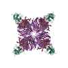
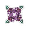
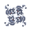
- PDB-1a14: COMPLEX BETWEEN NC10 ANTI-INFLUENZA VIRUS NEURAMINIDASE SINGLE CH... -
+ Open data

- Basic information
| Entry | Database: PDB / ID: 1a14 | |||||||||
|---|---|---|---|---|---|---|---|---|---|---|
| Title | COMPLEX BETWEEN NC10 ANTI-INFLUENZA VIRUS NEURAMINIDASE SINGLE CHAIN ANTIBODY WITH A 5 RESIDUE LINKER AND INFLUENZA VIRUS NEURAMINIDASE | |||||||||
 Components Components |
| |||||||||
 Keywords Keywords | COMPLEX (ANTIBODY/ANTIGEN) / COMPLEX (ANTIBODY-ANTIGEN) / SINGLE-CHAIN ANTIBODY / GLYCOSYLATED PROTEIN / COMPLEX (ANTIBODY-ANTIGEN) complex | |||||||||
| Function / homology |  Function and homology information Function and homology informationScavenging of heme from plasma / Fc epsilon receptor (FCERI) signaling / CD22 mediated BCR regulation / Role of LAT2/NTAL/LAB on calcium mobilization / FCERI mediated MAPK activation / Classical antibody-mediated complement activation / FCGR activation / Role of phospholipids in phagocytosis / Regulation of Complement cascade / FCERI mediated Ca+2 mobilization ...Scavenging of heme from plasma / Fc epsilon receptor (FCERI) signaling / CD22 mediated BCR regulation / Role of LAT2/NTAL/LAB on calcium mobilization / FCERI mediated MAPK activation / Classical antibody-mediated complement activation / FCGR activation / Role of phospholipids in phagocytosis / Regulation of Complement cascade / FCERI mediated Ca+2 mobilization / Cell surface interactions at the vascular wall / Antigen activates B Cell Receptor (BCR) leading to generation of second messengers / FCERI mediated NF-kB activation / Regulation of actin dynamics for phagocytic cup formation / Immunoregulatory interactions between a Lymphoid and a non-Lymphoid cell / exo-alpha-sialidase / immunoglobulin mediated immune response / exo-alpha-sialidase activity / immunoglobulin complex / antigen binding / viral budding from plasma membrane / carbohydrate metabolic process / adaptive immune response / immune response / host cell plasma membrane / virion membrane / extracellular region / membrane / metal ion binding / plasma membrane Similarity search - Function | |||||||||
| Biological species |    Influenza A virus Influenza A virus | |||||||||
| Method |  X-RAY DIFFRACTION / SYNCHROTRON / MOLECULAR REPLACEMENT / Resolution: 2.5 Å X-RAY DIFFRACTION / SYNCHROTRON / MOLECULAR REPLACEMENT / Resolution: 2.5 Å | |||||||||
 Authors Authors | Malby, R.L. / Mccoy, A.J. / Kortt, A.A. / Hudson, P.J. / Colman, P.M. | |||||||||
 Citation Citation | Journal: J.Mol.Biol. / Year: 1998 Title: Three-dimensional structures of single-chain Fv-neuraminidase complexes. Authors: Malby, R.L. / McCoy, A.J. / Kortt, A.A. / Hudson, P.J. / Colman, P.M. #1: Journal: Proteins / Year: 1993Title: Recombinant Antineuraminidase Single Chain Antibody: Expression, Characterization, and Crystallization in Complex with Antigen Authors: Malby, R.L. / Caldwell, J.B. / Gruen, L.C. / Harley, V.R. / Ivancic, N. / Kortt, A.A. / Lilley, G.G. / Power, B.E. / Webster, R.G. / Colman, P.M. / Hudson, P.J. | |||||||||
| History |
|
- Structure visualization
| Structure viewer | Molecule: MolmilJmol/JSmol |
|---|
- Downloads & links
-Download
| PDBx/mmCIF format | 1a14.cif.gz | 142.8 KB | Display | PDBx/mmCIF format |
|---|---|---|---|---|
| PDB format | pdb1a14.ent.gz | 109.2 KB | Display | PDB format |
| PDBx/mmJSON format | 1a14.json.gz | Tree view | PDBx/mmJSON format | |
| Others |  Other downloads Other downloads |
-Validation report
| Arichive directory | https://data.pdbj.org/pub/pdb/validation_reports/a1/1a14ftp://data.pdbj.org/pub/pdb/validation_reports/a1/1a14 | HTTPS FTP |
|---|
-Related structure data
| Related structure data |  1nmcSC S: Starting model for refinement C: citing same article ( |
|---|---|
| Similar structure data |

-Links
 PDBj
PDBj


- Assembly
| Deposited unit | 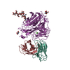
| ||||||||
|---|---|---|---|---|---|---|---|---|---|
| 1 | 
| ||||||||
| Unit cell |
|
-Components
-Antibody , 2 types, 2 molecules HL
| #2: Antibody | Mass: 13225.504 Da / Num. of mol.: 1 Fragment: VH DOMAIN OF ANTI-NEURAMINIDASE ANTIBODY NC10 COVALENTLY JOINED BY A FIVE-RESIDUE POLYPEPTIDE LINKER Source method: isolated from a genetically manipulated source Source: (gene. exp.)  |
|---|---|
| #3: Antibody | Mass: 11384.294 Da / Num. of mol.: 1 Fragment: VL DOMAIN OF ANTI-NEURAMINIDASE ANTIBODY NC10 COVALENTLY JOINED BY A FIVE-RESIDUE POLYPEPTIDE LINKER Source method: isolated from a genetically manipulated source Source: (gene. exp.) |
-Protein / Non-polymers , 2 types, 2 molecules N
| #1: Protein | Mass: 43723.770 Da / Num. of mol.: 1 / Fragment: RESIDUES 82 - 468 / Source method: isolated from a natural source / Source: (natural) Influenza A virus / Genus: Influenzavirus A / Strain: N9, A/TERN/AUSTRALIA/G70C/75 / References: UniProt: P03472, exo-alpha-sialidase |
|---|---|
| #7: Chemical | ChemComp-CA /  Mass: 40.078 Da / Num. of mol.: 1 / Source method: obtained synthetically / Formula: Ca Mass: 40.078 Da / Num. of mol.: 1 / Source method: obtained synthetically / Formula: Ca |
-Sugars , 3 types, 4 molecules 

| #4: Polysaccharide | alpha-D-mannopyranose-(1-2)-alpha-D-mannopyranose-(1-2)-alpha-D-mannopyranose-(1-3)-beta-D- ...alpha-D-mannopyranose-(1-2)-alpha-D-mannopyranose-(1-2)-alpha-D-mannopyranose-(1-3)-beta-D-mannopyranose-(1-4)-2-acetamido-2-deoxy-beta-D-glucopyranose-(1-4)-2-acetamido-2-deoxy-beta-D-glucopyranose Source method: isolated from a genetically manipulated source |
|---|---|
| #5: Sugar | ChemComp-MAN /  Type: D-saccharide, alpha linking / Mass: 180.156 Da / Num. of mol.: 1 Type: D-saccharide, alpha linking / Mass: 180.156 Da / Num. of mol.: 1Source method: isolated from a genetically manipulated source Formula: C6H12O6 |
| #6: Sugar |  Type: D-saccharide, beta linking / Mass: 221.208 Da / Num. of mol.: 2 Type: D-saccharide, beta linking / Mass: 221.208 Da / Num. of mol.: 2Source method: isolated from a genetically manipulated source Formula: C8H15NO6 |
-Details
| Has protein modification | Y |
|---|---|
| Nonpolymer details | THE THREE OLIGOSACCHARIDES COVALENTLY ATTACHED TO N9 NEURAMINIDASE ARE NUMBERED BY THE ASN RESIDUE ...THE THREE OLIGOSACCH |
-Experimental details
-Experiment
| Experiment | Method: X-RAY DIFFRACTION / Number of used crystals: 1 |
|---|
- Sample preparation
| Crystal | Density Matthews: 4 Å3/Da / Density % sol: 70 % | ||||||||||||||||||||||||||||||||||||
|---|---|---|---|---|---|---|---|---|---|---|---|---|---|---|---|---|---|---|---|---|---|---|---|---|---|---|---|---|---|---|---|---|---|---|---|---|---|
| Crystal grow | Method: vapor diffusion / pH: 6.6 Details: NC10 SCFV(5) AND N9 NA WERE MIXED TOGETHER (SCFV(5) IN FOUR-FOLD MOLAR EXCESS) WITH AND EQUAL VOLUME OF POTASSIUM PHOSPHATE BUFFER 1.7M PH6.6. THE DROP WAS EQUILIBRATED BY VAPOR DIFFUSION ...Details: NC10 SCFV(5) AND N9 NA WERE MIXED TOGETHER (SCFV(5) IN FOUR-FOLD MOLAR EXCESS) WITH AND EQUAL VOLUME OF POTASSIUM PHOSPHATE BUFFER 1.7M PH6.6. THE DROP WAS EQUILIBRATED BY VAPOR DIFFUSION AGAINST PHOSPHATE BUFFER 1.3M PH6.8., vapor diffusion PH range: 6.6-6.8 | ||||||||||||||||||||||||||||||||||||
| Crystal grow | *PLUS Method: vapor diffusion, sitting dropDetails: 1 was mixed with 2 in the molar ration 1:4, and an equal volume of 3 was added. | ||||||||||||||||||||||||||||||||||||
| Components of the solutions | *PLUS
|
-Data collection
| Diffraction | Mean temperature: 283 K |
|---|---|
| Diffraction source | Source: SYNCHROTRON / Site: Photon Factory  / Beamline: BL-18B / Wavelength: 1 / Beamline: BL-18B / Wavelength: 1 |
| Detector | Type: PHOTON FACTORY / Detector: IMAGE PLATE / Date: Jul 1, 1995 |
| Radiation | Monochromatic (M) / Laue (L): M / Scattering type: x-ray |
| Radiation wavelength | Wavelength: 1 Å / Relative weight: 1 |
| Reflection | Highest resolution: 2.5 Å / Num. obs: 29660 / % possible obs: 68 % / Observed criterion σ(I): 2 / Rmerge(I) obs: 0.118 |
| Reflection shell | Resolution: 2.5→2.6 Å / % possible all: 40 |
| Reflection | *PLUS Num. measured all: 86319 |
| Reflection shell | *PLUS % possible obs: 40 % |
- Processing
| Software |
| ||||||||||||||||||||||||||||||||||||||||||||||||||||||||||||
|---|---|---|---|---|---|---|---|---|---|---|---|---|---|---|---|---|---|---|---|---|---|---|---|---|---|---|---|---|---|---|---|---|---|---|---|---|---|---|---|---|---|---|---|---|---|---|---|---|---|---|---|---|---|---|---|---|---|---|---|---|---|
| Refinement | Method to determine structure: MOLECULAR REPLACEMENT Starting model: PDB ENTRY 1NMC Resolution: 2.5→7 Å / Data cutoff high absF: 10000000 / Data cutoff low absF: 0 / Cross valid method: A POSTERIORI / σ(F): 2
| ||||||||||||||||||||||||||||||||||||||||||||||||||||||||||||
| Refine analyze | Luzzati d res low obs: 5 Å | ||||||||||||||||||||||||||||||||||||||||||||||||||||||||||||
| Refinement step | Cycle: LAST / Resolution: 2.5→7 Å
| ||||||||||||||||||||||||||||||||||||||||||||||||||||||||||||
| Refine LS restraints |
| ||||||||||||||||||||||||||||||||||||||||||||||||||||||||||||
| Xplor file |
| ||||||||||||||||||||||||||||||||||||||||||||||||||||||||||||
| Software | *PLUS Name: X-PLOR / Version: 3.1 / Classification: refinement | ||||||||||||||||||||||||||||||||||||||||||||||||||||||||||||
| Refinement | *PLUS σ(I): 2 | ||||||||||||||||||||||||||||||||||||||||||||||||||||||||||||
| Solvent computation | *PLUS | ||||||||||||||||||||||||||||||||||||||||||||||||||||||||||||
| Displacement parameters | *PLUS |
