 Movie
Movie Controller


+ Open data

- Basic information
| Entry | Database: PDB / ID: 190d | ||||||||||||||||||
|---|---|---|---|---|---|---|---|---|---|---|---|---|---|---|---|---|---|---|---|
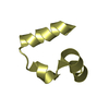
| Title | Crystal structure of a four-stranded intercalated DNA: d(C4) | ||||||||||||||||||
 Components Components | DNA (5'-D(* Keywords KeywordsDNA / U-DNA / QUADRUPLE HELIX / PARALLEL-STRANDED TETRAPLEX / BASE INTERCALATED / MISMATCHED | Function / homology | DNA |  Function and homology information Function and homology informationMethod |  X-RAY DIFFRACTION / Resolution: 1.8 Å X-RAY DIFFRACTION / Resolution: 1.8 Å  Authors AuthorsChen, L. / Cai, L. / Zhang, X. / Rich, A. |  CitationJournal: Biochemistry / Year: 1994 CitationJournal: Biochemistry / Year: 1994Title: Crystal structure of a four-stranded intercalated DNA: d(C4). Authors: Chen, L. / Cai, L. / Zhang, X. / Rich, A. History |
|
- Structure visualization
| Structure viewer | Molecule: MolmilJmol/JSmol |
|---|
- Downloads & links
-Download
| PDBx/mmCIF format | 190d.cif.gz | 33 KB | Display | PDBx/mmCIF format |
|---|---|---|---|---|
| PDB format | pdb190d.ent.gz | 24.8 KB | Display | PDB format |
| PDBx/mmJSON format | 190d.json.gz | Tree view | PDBx/mmJSON format | |
| Others |  Other downloads Other downloads |
-Validation report
| Arichive directory | https://data.pdbj.org/pub/pdb/validation_reports/90/190dftp://data.pdbj.org/pub/pdb/validation_reports/90/190d | HTTPS FTP |
|---|
-Related structure data
| Similar structure data |
|---|


-Links
 PDBj
PDBj

- Assembly
| Deposited unit | 
| ||||||||
|---|---|---|---|---|---|---|---|---|---|
| 1 | 
| ||||||||
| 2 | 
| ||||||||
| Unit cell |
|
-Components
| #1: DNA chain | Mass: 1111.770 Da / Num. of mol.: 8 / Source method: obtained synthetically #2: Water | ChemComp-HOH / |  Mass: 18.015 Da / Num. of mol.: 46 / Source method: isolated from a natural source / Formula: H2O Mass: 18.015 Da / Num. of mol.: 46 / Source method: isolated from a natural source / Formula: H2O |
|---|
-Experimental details
-Experiment
| Experiment | Method: X-RAY DIFFRACTION |
|---|
- Sample preparation
| Crystal | Density Matthews: 2.61 Å3/Da / Density % sol: 52.9 % | ||||||||||||||||||||||||||||||
|---|---|---|---|---|---|---|---|---|---|---|---|---|---|---|---|---|---|---|---|---|---|---|---|---|---|---|---|---|---|---|---|
| Crystal grow | Method: vapor diffusion, hanging drop / pH: 5.5 / Details: pH 5.50, VAPOR DIFFUSION, HANGING DROP | ||||||||||||||||||||||||||||||
| Components of the solutions |
| ||||||||||||||||||||||||||||||
| Crystal grow | *PLUS pH: 5.5 | ||||||||||||||||||||||||||||||
| Components of the solutions | *PLUS
|
-Data collection
| Diffraction | Ambient temp details: ROOM TEMPERATURE |
|---|---|
| Detector | Type: RIGAKU RAXIS II / Detector: IMAGE PLATE |
| Radiation | Scattering type: x-ray |
| Radiation wavelength | Relative weight: 1 |
- Processing
| Software | Name: X-PLOR / Classification: refinement | ||||||||||||||||||||||||||||||||||||||||||||||||||||||||||||
|---|---|---|---|---|---|---|---|---|---|---|---|---|---|---|---|---|---|---|---|---|---|---|---|---|---|---|---|---|---|---|---|---|---|---|---|---|---|---|---|---|---|---|---|---|---|---|---|---|---|---|---|---|---|---|---|---|---|---|---|---|---|
| Refinement | Resolution: 1.8→8 Å / σ(F): 2 Details: THIS IS THE FIRST X-RAY STRUCTURE OF THE INTERCALATED CYTOSINE QUADRUPLEX. THE PAPER CITED IN JRNL DESCRIBED THE D(C4) STRUCTURE REFINED AT 2.3 ANGSTROMS RESOLUTION. THE AUTHORS HAVE ...Details: THIS IS THE FIRST X-RAY STRUCTURE OF THE INTERCALATED CYTOSINE QUADRUPLEX. THE PAPER CITED IN JRNL DESCRIBED THE D(C4) STRUCTURE REFINED AT 2.3 ANGSTROMS RESOLUTION. THE AUTHORS HAVE EXTENDED THE STRUCTURE DETERMINATION TO 1.8 ANGSTROMS RESOLUTION AND THE COORDINATES DEPOSITED ARE THE FINAL 1.8 ANGSTROMS STRUCTURE WHICH IS BASICALLY THE SAME AS THAT AT 2.3 ANGSTROMS RESOLUTION. THERE ARE 8 D(C4) STRANDS PER ASYMMETRIC UNIT WHICH FORM TWO TETRAMERS. THE FIRST TETRAMER IS NUMBERED FROM 1 TO 16, AND THE OTHER TETRAMER FROM 17 TO 32. THERE ARE 46 WATER MOLECULES FOUND AT 1.8 A RESOLUTION. ALL HYDROGEN ATOMS ARE INCLUDED FOR EASY REFERENCE ALTHOUGH THEY ARE NOT REFINED WITH X-RAY DATA. THE STRUCTURE WAS SOLVED BY HEAVY ATOM PHASING. K2PTCL6 WAS SOAKED INTO THE CRYSTAL AND ONLY ONE PT SITE WAS FOUND WHICH WAS AT THE THREE-FOLD AXIS. SINGLE ISOMORPHOUS REPLACEMENT (SIR) PHASES AND SINGLE-WAVELENGTH ANOMALOUS SCATTERING (SAS) PHASES WERE IMPROVED BY SOLVENT FLATTENING. THE RESULTANT ELECTRON DENSITY MAP CLEARLY REVEALED THE INTERCALATED STRUCTURE FOR THE FIRST TIME.
| ||||||||||||||||||||||||||||||||||||||||||||||||||||||||||||
| Refine Biso |
| ||||||||||||||||||||||||||||||||||||||||||||||||||||||||||||
| Refinement step | Cycle: LAST / Resolution: 1.8→8 Å
| ||||||||||||||||||||||||||||||||||||||||||||||||||||||||||||
| Refine LS restraints |
| ||||||||||||||||||||||||||||||||||||||||||||||||||||||||||||
| Refinement | *PLUS Highest resolution: 2.3 Å / Lowest resolution: 8 Å / σ(F): 2 / Num. reflection obs: 4051 / Rfactor obs: 0.188 / Rfactor Rwork: 0.188 | ||||||||||||||||||||||||||||||||||||||||||||||||||||||||||||
| Solvent computation | *PLUS | ||||||||||||||||||||||||||||||||||||||||||||||||||||||||||||
| Displacement parameters | *PLUS | ||||||||||||||||||||||||||||||||||||||||||||||||||||||||||||
| Refine LS restraints | *PLUS
|
