 Movie
Movie Controller

[English] 日本語
 Yorodumi
- PDB-140d: SOLUTION STRUCTURE OF A CONSERVED DNA SEQUENCE FROM THE HIV-1 GEN... -
+ Open data

- Basic information
| Entry | Database: PDB / ID: 140d | ||||||
|---|---|---|---|---|---|---|---|
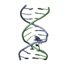
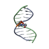
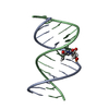
| Title | SOLUTION STRUCTURE OF A CONSERVED DNA SEQUENCE FROM THE HIV-1 GENOME: RESTRAINED MOLECULAR DYNAMICS SIMULATION WITH DISTANCE AND TORSION ANGLE RESTRAINTS DERIVED FROM TWO-DIMENSIONAL NMR SPECTRA | ||||||
 Components Components |
| ||||||
 Keywords Keywords | DNA / DOUBLE HELIX / CONSERVED SEQUENCE OF HIV-1 GENOME | ||||||
| Function / homology | DNA / DNA (> 10) Function and homology information Function and homology information | ||||||
| Method | SOLUTION NMR / MOLECULAR DYNAMICS, ENERGY MINIMIZATION | ||||||
 Authors Authors | Mujeeb, A. / Kerwin, S.M. / Kenyon, G.L. / James, T.L. | ||||||
 Citation Citation | Journal: Biochemistry / Year: 1993 Title: Solution structure of a conserved DNA sequence from the HIV-1 genome: restrained molecular dynamics simulation with distance and torsion angle restraints derived from two-dimensional NMR spectra. Authors: Mujeeb, A. / Kerwin, S.M. / Kenyon, G.L. / James, T.L. | ||||||
| History |
|
- Structure visualization
| Structure viewer | Molecule: MolmilJmol/JSmol |
|---|
- Downloads & links
-Download
| PDBx/mmCIF format | 140d.cif.gz | 20 KB | Display | PDBx/mmCIF format |
|---|---|---|---|---|
| PDB format | pdb140d.ent.gz | 12 KB | Display | PDB format |
| PDBx/mmJSON format | 140d.json.gz | Tree view | PDBx/mmJSON format | |
| Others |  Other downloads Other downloads |
-Validation report
| Summary document | 140d_validation.pdf.gz | 238.5 KB | Display | wwPDB validaton report |
|---|---|---|---|---|
| Full document | 140d_full_validation.pdf.gz | 238.2 KB | Display | |
| Data in XML | 140d_validation.xml.gz | 1.8 KB | Display | |
| Data in CIF | 140d_validation.cif.gz | 2 KB | Display | |
| Arichive directory | https://data.pdbj.org/pub/pdb/validation_reports/40/140dftp://data.pdbj.org/pub/pdb/validation_reports/40/140d | HTTPS FTP |
-Related structure data


-Links
 PDBj
PDBj


- Assembly
| Deposited unit | 
| |||||||||
|---|---|---|---|---|---|---|---|---|---|---|
| 1 |
| |||||||||
| NMR ensembles |
|
-Components
| #1: DNA chain | Mass: 3982.596 Da / Num. of mol.: 1 / Source method: obtained synthetically / Details: CHEMICALLY SYNTHESIZED |
|---|---|
| #2: DNA chain | Mass: 3960.600 Da / Num. of mol.: 1 / Source method: obtained synthetically / Details: CHEMICALLY SYNTHESIZED |
-Experimental details
-Experiment
| Experiment | Method: SOLUTION NMR |
|---|---|
| NMR details | Text: SUGAR PUCKER OF DEOXYRIBOSES HAS BEEN DETERMINED BY SIMULATION OF 2QF-COSY SPECTRA. A LIST OF TORSION ANGLE AND NOE DISTANCE RESTRAINTS IS AVAILABLE FROM THE PROTEIN DATA BANK AS ENTRY R140DMR. |
- Processing
| Software | Name:  AMBER / Classification: refinement AMBER / Classification: refinement | ||||||
|---|---|---|---|---|---|---|---|
| NMR software |
| ||||||
| Refinement | Method: MOLECULAR DYNAMICS, ENERGY MINIMIZATION / Software ordinal: 1 Details: ALL COORDINATES AROSE FROM ENERGY MINIMIZED AMBER4 FILES THAT WERE REFORMATTED FOR THE HELIX ANALYSIS PROGRAM CURVES. THEREFORE, ALL HYDROGEN ATOMS WERE REMOVED. THE DEPOSITORS HAVE PROVIDED ...Details: ALL COORDINATES AROSE FROM ENERGY MINIMIZED AMBER4 FILES THAT WERE REFORMATTED FOR THE HELIX ANALYSIS PROGRAM CURVES. THEREFORE, ALL HYDROGEN ATOMS WERE REMOVED. THE DEPOSITORS HAVE PROVIDED THREE COORDINATE SETS FOR THIS STRUCTURE. THE FIRST TWO COORDINATE SETS (PROTEIN DATA BANK ENTRIES 140D AND 141D) CONTAIN THE RESULTS OF THE NMR/RESTRAINED MOLECULAR DYNAMICS REFINEMENT WHERE A-DNA AND B-DNA WERE USED AS STARTING MODELS, RESPECTIVELY. THE AUTHORS DENOTED THESE STRUCTURES AS RMD-A AND RMD-B, RESPECTIVELY. THE THIRD COORDINATE SET (PROTEIN DATA BANK ENTRY 142D) REPRESENTS THE FINAL STRUCTURE, DENOTED RMD-FINAL BY THE AUTHORS. ALL STRUCTURES WERE DERIVED BY AVERAGING THE LAST 4 PS OF 30 PS RESTRAINED MD (AMBER4) AND SUBSEQUENT RESTRAINED ENERGY MINIMIZATION. FIVE RMD RUNS WERE AVERAGED TO EACH INTERIM STRUCTURE RMD-A AND RMD-B, DEPENDING ON THE STARTING GEOMETRY. ALL TEN STRUCTURES WERE AVERAGED, RESTRAINED ENERGY MINIMIZED AND A FINAL 20PS RMD RUN WAS PERFORMED, THE LAST 4PS OF WHICH, AFTER AVERAGING AND RESTRAINED ENERGY MINIMIZATION LEAD TO THE FINAL STRUCTURE: RMD-FINAL. | ||||||
| NMR ensemble | Conformers submitted total number: 1 |
