 Movie
Movie Controller


+ Open data

- Basic information
| Entry | Database: PDB / ID: 12zn | |||||||||
|---|---|---|---|---|---|---|---|---|---|---|
| Title | Septin tetrameric complex from Caenorhabditis elegans by Cryo-EM | |||||||||
 Components Components | (Septin) x 2 | |||||||||
 Keywords Keywords | CELL CYCLE / GTPase / Cytoskeleton component | |||||||||
| Function / homology |  Function and homology information Function and homology informationegg-laying behavior / septin complex / cytoskeleton-dependent cytokinesis / septin ring / post-embryonic development / locomotion / cell division site / cleavage furrow / intracellular protein localization / microtubule cytoskeleton ...egg-laying behavior / septin complex / cytoskeleton-dependent cytokinesis / septin ring / post-embryonic development / locomotion / cell division site / cleavage furrow / intracellular protein localization / microtubule cytoskeleton / midbody / molecular adaptor activity / cytoskeleton / GTPase activity / GTP binding Similarity search - Function | |||||||||
| Biological species |   Caenorhabditis elegans (invertebrata) Caenorhabditis elegans (invertebrata) | |||||||||
| Method | ELECTRON MICROSCOPY / single particle reconstruction / cryo EM / Resolution: 2.65 Å | |||||||||
 Authors Authors | Saladino, G.C.R. / Ciol, H. / Mendonca, D.C. / Furtado, A.A. / Pereira, H.M. / Klaholz, B. / Araujo, A.P.U. / Garratt, R.C. | |||||||||
| Funding support |  Brazil, 1items Brazil, 1items
| |||||||||
 Citation Citation | Journal: J Mol Biol / Year: 2026 Title: Cryo-EM Structure of the C. Elegans Septin Tetramer Reveals a Revised Architecture and Conserved Positional Orthology. Authors: Giovanna Christe Dos Reis Saladino / Heloísa Ciol / Deborah Cezar Mendonça / Adriano Alves Furtado / Humberto D'Muniz Pereira / Bruno P Klaholz / Ana Paula Ulian Araujo / Richard Charles Garratt /  Abstract: Septins are cytoskeletal proteins that assemble into hetero-oligomeric complexes, which polymerize into filaments to regulate many cellular processes, including cytokinesis and membrane remodeling. ...Septins are cytoskeletal proteins that assemble into hetero-oligomeric complexes, which polymerize into filaments to regulate many cellular processes, including cytokinesis and membrane remodeling. In Caenorhabditis elegans, the core septin complex is a tetramer composed of two copies each of two different subunits, UNC-59 and UNC-61. Historically, low-resolution models suggested a subunit order of UNC-59-UNC-61-UNC-61-UNC-59 for the tetramer. However, this arrangement contradicted the positional conservation of septins observed in other species. Using cryo-EM, we obtained a structure for the tetramer with a global resolution of 2.7 Å, definitively establishing the subunit order to be UNC-61-UNC-59-UNC-59-UNC-61. This places UNC-59 at the center of the particle where it forms a homodimeric G-interface, confirming it to be a positional ortholog of human SEPT7. This arrangement is further supported by mutational/biophysical analyses. Despite the stability of the tetramer, the complex failed to polymerize into filaments in vitro, which we attribute to the structural instability of the terminal UNC-61 G-interfaces. Our results suggest that post-translational modifications or specific cellular factors may be required to initiate higher-order assembly in C. elegans. Furthermore, our structure reveals that while UNC-59 possesses key characteristics of the SEPT7 group, it also features unique structural adaptations that may be related to the unique arrangement of the C. elegans complex. We demonstrate that the previous misidentification of the subunit order within the tetramer likely stemmed from N-terminal domain swapping, reinforcing the importance of high-resolution structural studies for understanding the evolutionary history and the diversity of septins. | |||||||||
| History |
|
- Structure visualization
| Structure viewer | Molecule: MolmilJmol/JSmol |
|---|
- Downloads & links
-Download
| PDBx/mmCIF format | 12zn.cif.gz | 245 KB | Display | PDBx/mmCIF format |
|---|---|---|---|---|
| PDB format | pdb12zn.ent.gz | 149.9 KB | Display | PDB format |
| PDBx/mmJSON format | 12zn.json.gz | Tree view | PDBx/mmJSON format | |
| Others |  Other downloads Other downloads |
-Validation report
| Arichive directory | https://data.pdbj.org/pub/pdb/validation_reports/2z/12znftp://data.pdbj.org/pub/pdb/validation_reports/2z/12zn | HTTPS FTP |
|---|
-Related structure data
| Related structure data |  76887MC M: map data used to model this data C: citing same article ( |
|---|---|
| Similar structure data |
-Links
 PDBj
PDBj- Assembly
| Deposited unit | 
|
|---|---|
| 1 |
|
-Components
| #1: Protein | Mass: 53135.703 Da / Num. of mol.: 2 Source method: isolated from a genetically manipulated source Source: (gene. exp.) Caenorhabditis elegans (invertebrata) / Gene: unc-61, CELE_Y50E8A.4, Y50E8A.4 / Plasmid: pETDuet / Production host:   Escherichia coli BL21(DE3) (bacteria) / Variant (production host): Rosetta / References: UniProt: Q9U277 Escherichia coli BL21(DE3) (bacteria) / Variant (production host): Rosetta / References: UniProt: Q9U277#2: Protein | Mass: 55299.492 Da / Num. of mol.: 2 Source method: isolated from a genetically manipulated source Source: (gene. exp.) Caenorhabditis elegans (invertebrata) / Gene: unc-59, CELE_W09C5.2, W09C5.2 / Plasmid: pETDuet / Production host: Escherichia coli BL21(DE3) (bacteria) / Variant (production host): Rosetta / References: UniProt: Q9U334#3: Chemical | 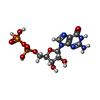 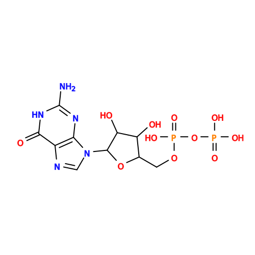 Type: RNA linking / Mass: 443.201 Da / Num. of mol.: 2 / Source method: obtained synthetically / Formula: C10H15N5O11P2 / Feature type: SUBJECT OF INVESTIGATION / Comment: GDP, energy-carrying molecule*YM Type: RNA linking / Mass: 443.201 Da / Num. of mol.: 2 / Source method: obtained synthetically / Formula: C10H15N5O11P2 / Feature type: SUBJECT OF INVESTIGATION / Comment: GDP, energy-carrying molecule*YMHas ligand of interest | Y | Has protein modification | N | |
|---|
-Experimental details
-Experiment
| Experiment | Method: ELECTRON MICROSCOPY |
|---|---|
| EM experiment | Aggregation state: PARTICLE / 3D reconstruction method: single particle reconstruction |
- Sample preparation
| Component | Name: Heterotetramer of C. elegans septins (UNC-61 - UNC-59 - UNC-59 - UNC-61) Type: COMPLEX / Entity ID: #1-#2 / Source: RECOMBINANT | ||||||||||||||||||||
|---|---|---|---|---|---|---|---|---|---|---|---|---|---|---|---|---|---|---|---|---|---|
| Molecular weight | Value: 0.20931 MDa / Experimental value: YES | ||||||||||||||||||||
| Source (natural) | Organism: Caenorhabditis elegans (invertebrata) | ||||||||||||||||||||
| Source (recombinant) | Organism: Escherichia coli (E. coli) / Strain: Rosetta DE3 / Plasmid: pETDuet | ||||||||||||||||||||
| Buffer solution | pH: 7.5 | ||||||||||||||||||||
| Buffer component |
| ||||||||||||||||||||
| Specimen | Conc.: 0.7 mg/ml / Embedding applied: NO / Shadowing applied: NO / Staining applied: NO / Vitrification applied: YES | ||||||||||||||||||||
| Specimen support | Grid material: GOLD / Grid mesh size: 300 divisions/in. / Grid type: Quantifoil R1.2/1.3 | ||||||||||||||||||||
| Vitrification | Instrument: FEI VITROBOT MARK IV / Cryogen name: ETHANE / Humidity: 100 % / Chamber temperature: 277 K |
- Electron microscopy imaging
| Experimental equipment |  Model: Titan Krios / Image courtesy: FEI Company |
|---|---|
| Microscopy | Model: TFS KRIOS |
| Electron gun | Electron source:  FIELD EMISSION GUN / Accelerating voltage: 300 kV / Illumination mode: FLOOD BEAM FIELD EMISSION GUN / Accelerating voltage: 300 kV / Illumination mode: FLOOD BEAM |
| Electron lens | Mode: BRIGHT FIELD / Nominal magnification: 165000 X / Nominal defocus max: 2000 nm / Nominal defocus min: 1500 nm / Cs: 2.7 mm |
| Specimen holder | Cryogen: NITROGEN / Specimen holder model: FEI TITAN KRIOS AUTOGRID HOLDER |
| Image recording | Electron dose: 44 e/Å2 / Film or detector model: FEI FALCON IV (4k x 4k) / Num. of real images: 5998 |
- Processing
| EM software |
| |||||||||||||||||||||||||||||||||||
|---|---|---|---|---|---|---|---|---|---|---|---|---|---|---|---|---|---|---|---|---|---|---|---|---|---|---|---|---|---|---|---|---|---|---|---|---|
| CTF correction | Type: NONE | |||||||||||||||||||||||||||||||||||
| Particle selection | Num. of particles selected: 5900000 | |||||||||||||||||||||||||||||||||||
| Symmetry | Point symmetry: C1 (asymmetric) | |||||||||||||||||||||||||||||||||||
| 3D reconstruction | Resolution: 2.65 Å / Resolution method: FSC 0.143 CUT-OFF / Num. of particles: 198470 / Symmetry type: POINT | |||||||||||||||||||||||||||||||||||
| Atomic model building | Protocol: RIGID BODY FIT / Space: REAL | |||||||||||||||||||||||||||||||||||
| Atomic model building |
| |||||||||||||||||||||||||||||||||||
| Refinement | Cross valid method: NONE Stereochemistry target values: GeoStd + Monomer Library + CDL v1.2 | |||||||||||||||||||||||||||||||||||
| Displacement parameters | Biso mean: 33.12 Å2 | |||||||||||||||||||||||||||||||||||
| Refine LS restraints |
|
