 Movie
Movie Controller

[English] 日本語
 Yorodumi
- PDB-12sl: Structure of a Strand-Displacement Complex of Human Mitochondrial... -
+ Open data

- Basic information
| Entry | Database: PDB / ID: 12sl | |||||||||
|---|---|---|---|---|---|---|---|---|---|---|
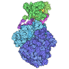
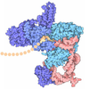
| Title | Structure of a Strand-Displacement Complex of Human Mitochondrial DNA Polymerase Gamma | |||||||||
 Components Components |
| |||||||||
 Keywords Keywords | REPLICATION / Mitochondrial DNA Polymerase Gamma / PolG / DNA strand displacement | |||||||||
| Function / homology |  Function and homology information Function and homology informationgamma DNA polymerase complex / mitochondrial chromosome / positive regulation of DNA-directed DNA polymerase activity / Strand-asynchronous mitochondrial DNA replication / mitochondrial DNA replication / DNA replication proofreading / single-stranded DNA 3'-5' DNA exonuclease activity / Hydrolases; Acting on ester bonds; Exodeoxyribonucleases producing 5'-phosphomonoesters / DNA metabolic process / DNA polymerase processivity factor activity ...gamma DNA polymerase complex / mitochondrial chromosome / positive regulation of DNA-directed DNA polymerase activity / Strand-asynchronous mitochondrial DNA replication / mitochondrial DNA replication / DNA replication proofreading / single-stranded DNA 3'-5' DNA exonuclease activity / Hydrolases; Acting on ester bonds; Exodeoxyribonucleases producing 5'-phosphomonoesters / DNA metabolic process / DNA polymerase processivity factor activity / Lyases; Carbon-oxygen lyases; Other carbon-oxygen lyases / mitochondrial nucleoid / 5'-deoxyribose-5-phosphate lyase activity / base-excision repair, gap-filling / DNA polymerase binding / 3'-5' exonuclease activity / DNA polymerase activity / Transcriptional activation of mitochondrial biogenesis / DNA-templated DNA replication / base-excision repair / protease binding / double-stranded DNA binding / DNA-directed DNA polymerase / DNA-directed DNA polymerase activity / mitochondrial matrix / chromatin binding / protein-containing complex / mitochondrion / DNA binding / identical protein binding Similarity search - Function | |||||||||
| Biological species |  Homo sapiens (human) Homo sapiens (human)synthetic construct (others) | |||||||||
| Method | ELECTRON MICROSCOPY / single particle reconstruction / cryo EM / Resolution: 2.93 Å | |||||||||
 Authors Authors | Nayak, A.R. / Sokolova, V.O. / Temiakov, D. | |||||||||
| Funding support |  United States, 1items United States, 1items
| |||||||||
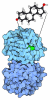
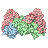
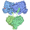
 Citation Citation | Journal: Nucleic Acids Res / Year: 2026 Title: Molecular and structural basis for replication initiation and strand separation by human mitochondrial DNA polymerase γ. Authors: Viktoriia Sokolova / Gina Buchel / Sarah Strock / Ashok R Nayak / Dmitry Temiakov / Abstract: Defects in human mitochondrial DNA (mtDNA) replication can lead to somatic mutations associated with a range of devastating mitochondrial diseases. However, the molecular mechanisms governing the ...Defects in human mitochondrial DNA (mtDNA) replication can lead to somatic mutations associated with a range of devastating mitochondrial diseases. However, the molecular mechanisms governing the earliest steps of mtDNA replication and their fidelity remain poorly understood. Here, we found that DNA polymerase gamma (Polγ) forms stable complexes with RNA-DNA primer-template substrates, exhibiting greater stability and lower misincorporation than on DNA-primed substrates. Structural analysis revealed that Polγ interacts with the 2'-OH groups of ribose within the first four nucleotides of the primer, explaining the stability of complexes that utilize RNA primers. Although Polγ requires TWINKLE to extend RNA primers, its intrinsic strand-displacement activity allows it to extend DNA primers independently. Structural data further show that the strand-separation mechanism in human Polγ is distinct from that of its yeast paralog, Mip1, and involves previously unresolved elements-the catcher and a GP loop in the exonuclease domain-that support intrinsic strand-displacement synthesis by Polγ. Structure-guided mutagenesis of elements involved in strand separation supports these structural observations. Together, our study provides mechanistic insight into mtDNA replication initiation and strand separation and has implications for understanding the molecular basis of mitochondrial disease. | |||||||||
| History |
|
- Structure visualization
| Structure viewer | Molecule: MolmilJmol/JSmol |
|---|
- Downloads & links
-Download
| PDBx/mmCIF format | 12sl.cif.gz | 385.5 KB | Display | PDBx/mmCIF format |
|---|---|---|---|---|
| PDB format | pdb12sl.ent.gz | 298.4 KB | Display | PDB format |
| PDBx/mmJSON format | 12sl.json.gz | Tree view | PDBx/mmJSON format | |
| Others |  Other downloads Other downloads |
-Validation report
| Arichive directory | https://data.pdbj.org/pub/pdb/validation_reports/2s/12slftp://data.pdbj.org/pub/pdb/validation_reports/2s/12sl | HTTPS FTP |
|---|
-Related structure data
| Related structure data |  76732MC  9pckC C: citing same article ( M: map data used to model this data |
|---|---|
| Similar structure data |


-Links
 PDBj
PDBj










- Assembly
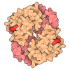
| Deposited unit | 
|
|---|---|
| 1 |
|
-Components
-DNA polymerase subunit gamma- ... , 2 types, 3 molecules ABC
| #1: Protein | Mass: 139730.703 Da / Num. of mol.: 1 Source method: isolated from a genetically manipulated source Source: (gene. exp.) Homo sapiens (human) / Gene: POLG, MDP1, POLG1, POLGA / Plasmid: pFastBac1 / Cell line (production host): Sf9 / Production host:   Spodoptera frugiperda (fall armyworm) / Tissue (production host): Ovary / References: UniProt: P54098, DNA-directed DNA polymerase Spodoptera frugiperda (fall armyworm) / Tissue (production host): Ovary / References: UniProt: P54098, DNA-directed DNA polymerase |
|---|---|
| #2: Protein | Mass: 54991.000 Da / Num. of mol.: 2 Source method: isolated from a genetically manipulated source Source: (gene. exp.) Homo sapiens (human) / Gene: POLG2, MTPOLB / Plasmid: pProEX / Production host:   Escherichia coli (E. coli) / Strain (production host): BL21 Codonplus (DE3) -RIPL / References: UniProt: Q9UHN1, DNA-directed DNA polymerase Escherichia coli (E. coli) / Strain (production host): BL21 Codonplus (DE3) -RIPL / References: UniProt: Q9UHN1, DNA-directed DNA polymerase |
-DNA chain , 2 types, 2 molecules PT
| #3: DNA chain | Mass: 5878.771 Da / Num. of mol.: 1 / Source method: obtained synthetically / Source: (synth.) synthetic construct (others) |
|---|---|
| #4: DNA chain | Mass: 7301.696 Da / Num. of mol.: 1 / Source method: obtained synthetically / Source: (synth.) synthetic construct (others) |
-Non-polymers , 3 types, 3 molecules 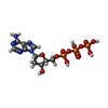

| #5: Chemical | ChemComp-DTP / 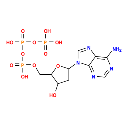 Mass: 491.182 Da / Num. of mol.: 1 / Source method: obtained synthetically / Formula: C10H16N5O12P3 Mass: 491.182 Da / Num. of mol.: 1 / Source method: obtained synthetically / Formula: C10H16N5O12P3 |
|---|---|
| #6: Chemical | ChemComp-MG /  Mass: 24.305 Da / Num. of mol.: 1 / Source method: obtained synthetically / Formula: Mg Mass: 24.305 Da / Num. of mol.: 1 / Source method: obtained synthetically / Formula: Mg |
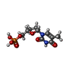
| #7: Chemical | ChemComp-2DT / 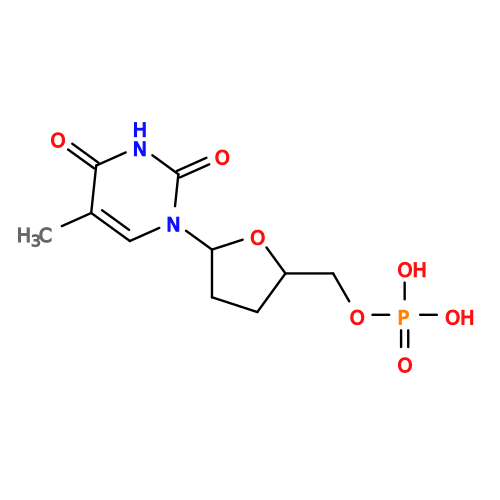 Type: DNA linking / Mass: 306.209 Da / Num. of mol.: 1 / Source method: obtained synthetically / Formula: C10H15N2O7P Type: DNA linking / Mass: 306.209 Da / Num. of mol.: 1 / Source method: obtained synthetically / Formula: C10H15N2O7P |
-Details
| Has ligand of interest | N |
|---|---|
| Has protein modification | Y |
-Experimental details
-Experiment
| Experiment | Method: ELECTRON MICROSCOPY |
|---|---|
| EM experiment | Aggregation state: PARTICLE / 3D reconstruction method: single particle reconstruction |
- Sample preparation
| Component | Name: Strand displacement Complex of Human Mitochondrial DNA Polymerase Gamma Type: COMPLEX Details: Strand-displacement complex of human mitochondrial DNA polymerase Gamma (exo-), assembled on a 1-nt gapped primer-template substrate contiguous with a downstream duplex containing a 4-nt 5' ...Details: Strand-displacement complex of human mitochondrial DNA polymerase Gamma (exo-), assembled on a 1-nt gapped primer-template substrate contiguous with a downstream duplex containing a 4-nt 5' non-template flap ss-DNA. The primer terminus was extended by four nucleotides (three dG followed by a chain-terminating ddT), thereby promoting coordinated displacement of four non-template nucleotides from the downstream DNA duplex. The ternary catalytic complex is stabilized by a bound incoming nucleotide (dATP) at the polymerase active site in the presence of 5 mM MgCl2. Entity ID: #1-#4 / Source: RECOMBINANT |
|---|---|
| Molecular weight | Value: 0.362 MDa / Experimental value: YES |
| Source (natural) | Organism: Homo sapiens (human) |
| Source (recombinant) | Organism: Spodoptera frugiperda (fall armyworm) |
| Buffer solution | pH: 7.9 Details: 10 mM Tris-Hcl pH 7.9, 100 mM Nacl, 10 mM DTT, and 5 mM MgCl2 |
| Specimen | Conc.: 0.52 mg/ml / Embedding applied: NO / Shadowing applied: NO / Staining applied: NO / Vitrification applied: YES Details: 2 uM strand-displacement complex of human mitochondrial DNA polymerase Gamma (exo-), assembled on an equimolar 1-nt gapped primer-template substrate contiguous with a downstream duplex ...Details: 2 uM strand-displacement complex of human mitochondrial DNA polymerase Gamma (exo-), assembled on an equimolar 1-nt gapped primer-template substrate contiguous with a downstream duplex containing a 4-nt single-stranded non-template flap. The primer was extended by four nucleotides; three successive dG at 0.2 mM, followed by incorporation of a chain-terminating ddT at 0.1 mM), thereby driving coordinated displacement of four nucleotides from the downstream DNA duplex. The resulting ternary catalytic complex was further stabilized by binding of an incoming nucleotide (1 mM dATP) at the polymerase active site in the presence of 5 mM MgCl2. |
| Specimen support | Grid material: GOLD / Grid mesh size: 300 divisions/in. / Grid type: UltrAuFoil R1.2/1.3 |
| Vitrification | Instrument: FEI VITROBOT MARK IV / Cryogen name: ETHANE / Humidity: 100 % / Chamber temperature: 277 K |
- Electron microscopy imaging
| Experimental equipment |  Model: Titan Krios / Image courtesy: FEI Company |
|---|---|
| Microscopy | Model: TFS KRIOS |
| Electron gun | Electron source:  FIELD EMISSION GUN / Accelerating voltage: 300 kV / Illumination mode: FLOOD BEAM FIELD EMISSION GUN / Accelerating voltage: 300 kV / Illumination mode: FLOOD BEAM |
| Electron lens | Mode: BRIGHT FIELD / Nominal magnification: 105000 X / Nominal defocus max: 1500 nm / Nominal defocus min: 500 nm / Cs: 2.7 mm / C2 aperture diameter: 100 µm / Alignment procedure: COMA FREE |
| Specimen holder | Cryogen: NITROGEN |
| Image recording | Average exposure time: 2.6 sec. / Electron dose: 60 e/Å2 / Film or detector model: GATAN K3 (6k x 4k) / Num. of grids imaged: 1 / Num. of real images: 7236 |
| EM imaging optics | Energyfilter slit width: 20 eV |
- Processing
| EM software |
| ||||||||||||||||||||||||||||||||
|---|---|---|---|---|---|---|---|---|---|---|---|---|---|---|---|---|---|---|---|---|---|---|---|---|---|---|---|---|---|---|---|---|---|
| CTF correction | Type: PHASE FLIPPING AND AMPLITUDE CORRECTION | ||||||||||||||||||||||||||||||||
| Particle selection | Num. of particles selected: 5827907 | ||||||||||||||||||||||||||||||||
| Symmetry | Point symmetry: C1 (asymmetric) | ||||||||||||||||||||||||||||||||
| 3D reconstruction | Resolution: 2.93 Å / Resolution method: FSC 0.143 CUT-OFF / Num. of particles: 395220 / Num. of class averages: 1 / Symmetry type: POINT | ||||||||||||||||||||||||||||||||
| Atomic model building | Protocol: FLEXIBLE FIT / Space: REAL | ||||||||||||||||||||||||||||||||
| Atomic model building | PDB-ID: 9PCK Accession code: 9PCK / Source name: PDB / Type: experimental model |
