 Movie
Movie Controller

[English] 日本語
 Yorodumi
- EMDB-77235: Focused map (Accessory Subunit) of the Strand-Displacement Comple... -
+ Open data

- Basic information
| Entry |  | |||||||||
|---|---|---|---|---|---|---|---|---|---|---|
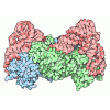
| Title | Focused map (Accessory Subunit) of the Strand-Displacement Complex of Human Mitochondrial DNA Polymerase Gamma | |||||||||
 Map data Map data | ||||||||||
 Sample Sample |
| |||||||||
 Keywords Keywords | Mitochondrial DNA Polymerase Gamma / PolG / DNA strand displacement / REPLICATION | |||||||||
| Function / homology |  Function and homology information Function and homology informationgamma DNA polymerase complex / positive regulation of DNA-directed DNA polymerase activity / Strand-asynchronous mitochondrial DNA replication / mitochondrial DNA replication / DNA polymerase processivity factor activity / mitochondrial nucleoid / DNA polymerase binding / Transcriptional activation of mitochondrial biogenesis / DNA-templated DNA replication / double-stranded DNA binding ...gamma DNA polymerase complex / positive regulation of DNA-directed DNA polymerase activity / Strand-asynchronous mitochondrial DNA replication / mitochondrial DNA replication / DNA polymerase processivity factor activity / mitochondrial nucleoid / DNA polymerase binding / Transcriptional activation of mitochondrial biogenesis / DNA-templated DNA replication / double-stranded DNA binding / DNA-directed DNA polymerase activity / mitochondrial matrix / mitochondrion / identical protein binding Similarity search - Function | |||||||||
| Biological species |  Homo sapiens (human) Homo sapiens (human) | |||||||||
| Method | single particle reconstruction / cryo EM / Resolution: 2.81 Å | |||||||||
 Authors Authors | Nayak AR / Sokolova VO / Temiakov D | |||||||||
| Funding support |  United States, 1 items United States, 1 items
| |||||||||
 Citation Citation | Journal: Nucleic Acids Res / Year: 2026 Title: Molecular and structural basis for replication initiation and strand separation by human mitochondrial DNA polymerase γ. Authors: Viktoriia Sokolova / Gina Buchel / Sarah Strock / Ashok R Nayak / Dmitry Temiakov / Abstract: Defects in human mitochondrial DNA (mtDNA) replication can lead to somatic mutations associated with a range of devastating mitochondrial diseases. However, the molecular mechanisms governing the ...Defects in human mitochondrial DNA (mtDNA) replication can lead to somatic mutations associated with a range of devastating mitochondrial diseases. However, the molecular mechanisms governing the earliest steps of mtDNA replication and their fidelity remain poorly understood. Here, we found that DNA polymerase gamma (Polγ) forms stable complexes with RNA-DNA primer-template substrates, exhibiting greater stability and lower misincorporation than on DNA-primed substrates. Structural analysis revealed that Polγ interacts with the 2'-OH groups of ribose within the first four nucleotides of the primer, explaining the stability of complexes that utilize RNA primers. Although Polγ requires TWINKLE to extend RNA primers, its intrinsic strand-displacement activity allows it to extend DNA primers independently. Structural data further show that the strand-separation mechanism in human Polγ is distinct from that of its yeast paralog, Mip1, and involves previously unresolved elements-the catcher and a GP loop in the exonuclease domain-that support intrinsic strand-displacement synthesis by Polγ. Structure-guided mutagenesis of elements involved in strand separation supports these structural observations. Together, our study provides mechanistic insight into mtDNA replication initiation and strand separation and has implications for understanding the molecular basis of mitochondrial disease. | |||||||||
| History |
|
- Structure visualization
| Supplemental images |
|---|

- Downloads & links
-EMDB archive
| Map data | emd_77235.map.gz | 7.6 MB | EMDB map data format | |
|---|---|---|---|---|
| Header (meta data) | emd-77235-v30.xmlemd-77235.xml | 22 KB 22 KB | Display Display | EMDB header |
| FSC (resolution estimation) | emd_77235_fsc.xml | 10.7 KB | Display | FSC data file |
| Images |  emd_77235.png emd_77235.png | 118.3 KB | ||
| Masks | emd_77235_msk_1.map | 129.7 MB | Mask map | |
| Filedesc metadata | emd-77235.cif.gz | 6.2 KB | ||
| Others | emd_77235_additional_1.map.gzemd_77235_half_map_1.map.gzemd_77235_half_map_2.map.gz | 65.1 MB 120.3 MB 120.3 MB | ||
| Archive directory |  https://data.pdbj.org/pub/emdb/structures/EMD-77235ftp://data.pdbj.org/pub/emdb/structures/EMD-77235 https://data.pdbj.org/pub/emdb/structures/EMD-77235ftp://data.pdbj.org/pub/emdb/structures/EMD-77235 | HTTPS FTP |
-Related structure data


-Links
| EMDB pages | EMDB (EBI/PDBe) / EMDataResource |
|---|---|
| Related items in Molecule of the Month |
-Map
| File | Download / File: emd_77235.map.gz / Format: CCP4 / Size: 129.7 MB / Type: IMAGE STORED AS FLOATING POINT NUMBER (4 BYTES) | ||||||||||||||||||||||||||||||||||||
|---|---|---|---|---|---|---|---|---|---|---|---|---|---|---|---|---|---|---|---|---|---|---|---|---|---|---|---|---|---|---|---|---|---|---|---|---|---|
| Projections & slices | Image control
Images are generated by Spider. | ||||||||||||||||||||||||||||||||||||
| Voxel size | X=Y=Z: 0.8556 Å | ||||||||||||||||||||||||||||||||||||
| Density |
| ||||||||||||||||||||||||||||||||||||
| Symmetry | Space group: 1 | ||||||||||||||||||||||||||||||||||||
| Details | EMDB XML:
|
 Z (Sec.)
 Y (Row.)
 X (Col.)


-Supplemental data
-Mask #1
| File | emd_77235_msk_1.map | ||||||||||||
|---|---|---|---|---|---|---|---|---|---|---|---|---|---|
| Projections & Slices |
| ||||||||||||
| Density Histograms |


-Additional map: #1
| File | emd_77235_additional_1.map | ||||||||||||
|---|---|---|---|---|---|---|---|---|---|---|---|---|---|
| Projections & Slices |
| ||||||||||||
| Density Histograms |


-Half map: #1
| File | emd_77235_half_map_1.map | ||||||||||||
|---|---|---|---|---|---|---|---|---|---|---|---|---|---|
| Projections & Slices |
| ||||||||||||
| Density Histograms |


-Half map: #2
| File | emd_77235_half_map_2.map | ||||||||||||
|---|---|---|---|---|---|---|---|---|---|---|---|---|---|
| Projections & Slices |
| ||||||||||||
| Density Histograms |


- Sample components
-Entire : Strand displacement Complex of Human Mitochondrial DNA Polymerase...
| Entire | Name: Strand displacement Complex of Human Mitochondrial DNA Polymerase Gamma |
|---|---|
| Components |
|
-Supramolecule #1: Strand displacement Complex of Human Mitochondrial DNA Polymerase...
| Supramolecule | Name: Strand displacement Complex of Human Mitochondrial DNA Polymerase Gamma type: complex / ID: 1 / Parent: 0 / Macromolecule list: all Details: Strand-displacement complex of human mitochondrial DNA polymerase Gamma (exo-), assembled on a 1-nt gapped primer-template substrate contiguous with a downstream duplex containing a 4-nt 5' ...Details: Strand-displacement complex of human mitochondrial DNA polymerase Gamma (exo-), assembled on a 1-nt gapped primer-template substrate contiguous with a downstream duplex containing a 4-nt 5' non-template flap ss-DNA. The primer terminus was extended by four nucleotides (three dG followed by a chain-terminating ddT), thereby promoting coordinated displacement of four non-template nucleotides from the downstream DNA duplex. The ternary catalytic complex is stabilized by a bound incoming nucleotide (dATP) at the polymerase active site in the presence of 5 mM MgCl2. |
|---|---|
| Source (natural) | Organism: Homo sapiens (human) |
| Molecular weight | Theoretical: 362 KDa |
-Macromolecule #1: DNA polymerase subunit gamma-2, mitochondrial
| Macromolecule | Name: DNA polymerase subunit gamma-2, mitochondrial / type: protein_or_peptide / ID: 1 / Details: Accessory subunit / Enantiomer: LEVO / EC number: DNA-directed DNA polymerase |
|---|---|
| Source (natural) | Organism: Homo sapiens (human) |
| Recombinant expression | Organism:   Escherichia coli (E. coli) Escherichia coli (E. coli) |
| Sequence | String: MRSRVAVRAC HKVCRCLLSG FGGRVDAGQP ELLTERSSPK GGHVKSHAEL EGNGEHPEAP GSGEGSEALL EICQRRHFLS GSKQQLSRD SLLSGCHPGF GPLGVELRKN LAAEWWTSVV VFREQVFPVD ALHHKPGPLL PGDSAFRLVS AETLREILQD K ELSKEQLV ...String: MRSRVAVRAC HKVCRCLLSG FGGRVDAGQP ELLTERSSPK GGHVKSHAEL EGNGEHPEAP GSGEGSEALL EICQRRHFLS GSKQQLSRD SLLSGCHPGF GPLGVELRKN LAAEWWTSVV VFREQVFPVD ALHHKPGPLL PGDSAFRLVS AETLREILQD K ELSKEQLV AFLENVLKTS GKLRENLLHG ALEHYVNCLD LVNKRLPYGL AQIGVCFHPV FDTKQIRNGV KSIGEKTEAS LV WFTPPRT SNQWLDFWLR HRLQWWRKFA MSPSNFSSSD CQDEEGRKGN KLYYNFPWGK ELIETLWNLG DHELLHMYPG NVS KLHGRD GRKNVVPCVL SVNGDLDRGM LAYLYDSFQL TENSFTRKKN LHRKVLKLHP CLAPIKVALD VGRGPTLELR QVCQ GLFNE LLENGISVWP GYLETMQSSL EQLYSKYDEM SILFTVLVTE TTLENGLIHL RSRDTTMKEM MHISKLKDFL IKYIS SAKN V UniProtKB: DNA polymerase subunit gamma-2 |
-Experimental details
-Structure determination
| Method | cryo EM |
|---|---|
 Processing Processing | single particle reconstruction |
| Aggregation state | particle |
-Sample preparation
| Concentration | 0.52 mg/mL |
|---|---|
| Buffer | pH: 7.9 Details: 10 mM Tris-Hcl pH 7.9, 100 mM Nacl, 10 mM DTT, and 5 mM MgCl2 |
| Grid | Model: UltrAuFoil R1.2/1.3 / Material: GOLD / Pretreatment - Type: GLOW DISCHARGE / Pretreatment - Atmosphere: AIR |
| Vitrification | Cryogen name: ETHANE / Chamber humidity: 100 % / Chamber temperature: 277 K / Instrument: FEI VITROBOT MARK IV |
| Details | 2 uM strand-displacement complex of human mitochondrial DNA polymerase Gamma (exo-), assembled on an equimolar 1-nt gapped primer-template substrate contiguous with a downstream duplex containing a 4-nt single-stranded non-template flap. The primer was extended by four nucleotides; three successive dG at 0.2 mM, followed by incorporation of a chain-terminating ddT at 0.1 mM), thereby driving coordinated displacement of four nucleotides from the downstream DNA duplex. The resulting ternary catalytic complex was further stabilized by binding of an incoming nucleotide (1 mM dATP) at the polymerase active site in the presence of 5 mM MgCl2. |
- Electron microscopy
| Microscope | TFS KRIOS |
|---|---|
| Specialist optics | Energy filter - Slit width: 20 eV |
| Software | Name: Latitude |
| Image recording | Film or detector model: GATAN K3 (6k x 4k) / Number grids imaged: 1 / Number real images: 7236 / Average exposure time: 2.6 sec. / Average electron dose: 60.0 e/Å2 |
| Electron beam | Acceleration voltage: 300 kV / Electron source:  FIELD EMISSION GUN FIELD EMISSION GUN |
| Electron optics | C2 aperture diameter: 100.0 µm / Illumination mode: FLOOD BEAM / Imaging mode: BRIGHT FIELD / Cs: 2.7 mm / Nominal defocus max: 1.5 µm / Nominal defocus min: 0.5 µm / Nominal magnification: 105000 |
| Sample stage | Cooling holder cryogen: NITROGEN |
| Experimental equipment |  Model: Titan Krios / Image courtesy: FEI Company |
+Image processing

-Atomic model buiding 1
| Initial model | PDB ID: Chain - Source name: PDB / Chain - Initial model type: experimental model |
|---|---|
| Software | Name: Coot (ver. 0.9.8.5) |
| Refinement | Space: REAL / Protocol: FLEXIBLE FIT |
