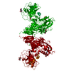
Movie
Movie Controller

[English] 日本語
 Yorodumi
- PDB-12bn: Structural insights into the exosite-mediated activation of Facto... -
+ Open data

- Basic information
| Entry | Database: PDB / ID: 12bn | ||||||||||||||||||
|---|---|---|---|---|---|---|---|---|---|---|---|---|---|---|---|---|---|---|---|
| Title | Structural insights into the exosite-mediated activation of Factor IX by Factor XIa using cryoEM | ||||||||||||||||||
 Components Components |
| ||||||||||||||||||
 Keywords Keywords | BLOOD CLOTTING / Coagulation / Intrinsic Pathway / Complex / Hemophilia / Factor IX / Factor XIa | ||||||||||||||||||
| Function / homology |  Function and homology information Function and homology informationDefective F9 secretion / coagulation factor IXa / Defective gamma-carboxylation of F9 / coagulation factor XIa / serine-type aminopeptidase activity / Defective F9 activation / positive regulation of fibrinolysis / Defective factor IX causes thrombophilia / Defective cofactor function of FVIIIa variant / Defective F9 variant does not activate FX ...Defective F9 secretion / coagulation factor IXa / Defective gamma-carboxylation of F9 / coagulation factor XIa / serine-type aminopeptidase activity / Defective F9 activation / positive regulation of fibrinolysis / Defective factor IX causes thrombophilia / Defective cofactor function of FVIIIa variant / Defective F9 variant does not activate FX / : / zymogen activation / plasminogen activation / Protein hydroxylation / Transport of gamma-carboxylated protein precursors from the endoplasmic reticulum to the Golgi apparatus / Gamma-carboxylation of protein precursors / Removal of aminoterminal propeptides from gamma-carboxylated proteins / : / serine-type peptidase activity / Golgi lumen / blood coagulation / heparin binding / endopeptidase activity / extracellular matrix / endoplasmic reticulum lumen / serine-type endopeptidase activity / calcium ion binding / proteolysis / : / extracellular exosome / extracellular region / membrane / metal ion binding / identical protein binding / plasma membrane Similarity search - Function | ||||||||||||||||||
| Biological species |  Homo sapiens (human) Homo sapiens (human) | ||||||||||||||||||
| Method | ELECTRON MICROSCOPY / single particle reconstruction / cryo EM / Resolution: 3.4 Å | ||||||||||||||||||
 Authors Authors | Mohammed, B.M. | ||||||||||||||||||
| Funding support |  United States, 5items United States, 5items
| ||||||||||||||||||
 Citation Citation | Journal: J Thromb Haemost / Year: 2026 Title: Structural insights into the exosite-mediated activation of Factor IX by Factor XIa using cryo-EM. Authors: Bassem M Mohammed / Samantha Deavila / Tristan Friet / Isabella Dattilio / Abstract: BACKGROUND: Factor XI (FXI) occupies a unique and clinically significant niche, bridging the tissue factor-driven and contact-driven coagulation pathways. Irrespective of the trigger, activated ...BACKGROUND: Factor XI (FXI) occupies a unique and clinically significant niche, bridging the tissue factor-driven and contact-driven coagulation pathways. Irrespective of the trigger, activated factor XI (FXIa) contributes to clotting by activating factor IX (FIX). Biochemical studies established that this reaction requires the membrane-binding FIX-Gla domain to engage an exosite on the FXIa Apple 3 (A3) domain, that only become available upon FXI activation. Structural data for FXIa, FIX, FIXaβ, and the FXIa:FIX complex are lacking; current understanding relies on zymogen FXI crystal structures and homology modeling of FXIa after kallikrein. OBJECTIVES: Elucidate the high-resolution structure of FXIa in functionally relevant conformation in complex with FIX. METHODS: We utilized cryogenic electron microscopy (cryo-EM) to determine the structures of human FXIa in complex with its full-length substrate, FIX, and activated product, FIXaβ. RESULTS: We report the first cryo-EM structures of FXIa in complex with its substrate, FIX, and activated FIX (FIXaβ). The structures capture a functionally relevant conformational change in the ...RESULTS: We report the first cryo-EM structures of FXIa in complex with its substrate, FIX, and activated FIX (FIXaβ). The structures capture a functionally relevant conformational change in the FXIa catalytic domain and reveals the first view of the entire FIX and FIXaβ. Critically, we visualize the FIX-Gla domain precisely docked to the FXIa-A3 exosite on both subunits of the FXIa dimer. We also define the first step of proteolysis, visualizing the FIX Arg145 inserted into the primary specificity pocket of FXIa. CONCLUSIONS: The structures define the full FXIa:FIX interface providing a structural template for understanding the sequential activation of FIX and for developing a new class of selective ...CONCLUSIONS: The structures define the full FXIa:FIX interface providing a structural template for understanding the sequential activation of FIX and for developing a new class of selective allosteric antithrombotic agents. | ||||||||||||||||||
| History |
|
- Structure visualization
| Structure viewer | Molecule: MolmilJmol/JSmol |
|---|
- Downloads & links
-Download
| PDBx/mmCIF format | 12bn.cif.gz | 337.2 KB | Display | PDBx/mmCIF format |
|---|---|---|---|---|
| PDB format | pdb12bn.ent.gz | 267.8 KB | Display | PDB format |
| PDBx/mmJSON format | 12bn.json.gz | Tree view | PDBx/mmJSON format | |
| Others |  Other downloads Other downloads |
-Validation report
| Arichive directory | https://data.pdbj.org/pub/pdb/validation_reports/2b/12bnftp://data.pdbj.org/pub/pdb/validation_reports/2b/12bn | HTTPS FTP |
|---|
-Related structure data
| Related structure data |  76288MC  9zqyC  9ztkC  9zubC  9zunC  9zvxC C: citing same article ( M: map data used to model this data |
|---|---|
| Similar structure data |


-Links
 PDBj
PDBj


- Assembly
| Deposited unit | 
|
|---|---|
| 1 |
|
-Components
| #1: Protein | Mass: 47158.219 Da / Num. of mol.: 2 / Source method: isolated from a natural source / Source: (natural) Homo sapiens (human) / Tissue: plasma / References: UniProt: P00740, coagulation factor IXa#2: Protein | Mass: 41262.098 Da / Num. of mol.: 2 Source method: isolated from a genetically manipulated source Details: Activated Recombinant Factor XI Ser557Ala. The molecule is a dimer that is disulphide linked at Cys321. During activation the bond at R369 is cleaved on each subunit to give a two chain ...Details: Activated Recombinant Factor XI Ser557Ala. The molecule is a dimer that is disulphide linked at Cys321. During activation the bond at R369 is cleaved on each subunit to give a two chain molecule. only one monomer is solved here and is composed of a heavy chain and light chain Source: (gene. exp.) Homo sapiens (human) / Gene: F11 / Plasmid: pDest40 / Cell line (production host): HEK 293 cells / Production host: Homo sapiens (human) / References: UniProt: P03951, coagulation factor XIa#3: Antibody | Mass: 26686.352 Da / Num. of mol.: 2 / Mutation: S557A Source method: isolated from a genetically manipulated source Details: Has a Ser557Ala mutation to render it catalytically dead. The molecule is a dimer that is disulphide linked at Cys321. During activation the bond at R369 is cleaved on each subunit to give a ...Details: Has a Ser557Ala mutation to render it catalytically dead. The molecule is a dimer that is disulphide linked at Cys321. During activation the bond at R369 is cleaved on each subunit to give a two chain molecule. only one monomer is solved here and is composed of a heavy chain and light chain Source: (gene. exp.) Homo sapiens (human) / Gene: F11 / Plasmid: pDest40 / Cell line (production host): HEK 293 cells / Production host: Homo sapiens (human) / References: UniProt: P03951#4: Chemical | ChemComp-CA /   Mass: 40.078 Da / Num. of mol.: 16 / Source method: obtained synthetically / Formula: Ca / Feature type: SUBJECT OF INVESTIGATION Mass: 40.078 Da / Num. of mol.: 16 / Source method: obtained synthetically / Formula: Ca / Feature type: SUBJECT OF INVESTIGATION#5: Sugar | ChemComp-NAG /   Type: D-saccharide, beta linking / Mass: 221.208 Da / Num. of mol.: 8 Type: D-saccharide, beta linking / Mass: 221.208 Da / Num. of mol.: 8Source method: isolated from a genetically manipulated source Formula: C8H15NO6 / Feature type: SUBJECT OF INVESTIGATION Has ligand of interest | Y | Has protein modification | Y | |
|---|
-Experimental details
-Experiment
| Experiment | Method: ELECTRON MICROSCOPY |
|---|---|
| EM experiment | Aggregation state: PARTICLE / 3D reconstruction method: single particle reconstruction |
- Sample preparation
| Component |
| |||||||||||||||||||||||||||||||||||
|---|---|---|---|---|---|---|---|---|---|---|---|---|---|---|---|---|---|---|---|---|---|---|---|---|---|---|---|---|---|---|---|---|---|---|---|---|
| Molecular weight |
| |||||||||||||||||||||||||||||||||||
| Source (natural) |
| |||||||||||||||||||||||||||||||||||
| Source (recombinant) | Organism: Homo sapiens (human) / Cell: HEK293 / Plasmid: pDest40 | |||||||||||||||||||||||||||||||||||
| Buffer solution |
| |||||||||||||||||||||||||||||||||||
| Buffer component |
| |||||||||||||||||||||||||||||||||||
| Specimen |
| |||||||||||||||||||||||||||||||||||
| Specimen support |
| |||||||||||||||||||||||||||||||||||
| Vitrification | Chamber temperature: 277.15 K / Cryogen name: ETHANE / Entry-ID: 12BN / Humidity: 95 % / Instrument: FEI VITROBOT MARK IV
|
- Electron microscopy imaging
| Microscopy | Model: TFS GLACIOS | |||||||||||||||||||||
|---|---|---|---|---|---|---|---|---|---|---|---|---|---|---|---|---|---|---|---|---|---|---|
| Electron gun | Electron source:  FIELD EMISSION GUN / Accelerating voltage: 200 kV / Illumination mode: FLOOD BEAM FIELD EMISSION GUN / Accelerating voltage: 200 kV / Illumination mode: FLOOD BEAM | |||||||||||||||||||||
| Electron lens | Mode: BRIGHT FIELD / Nominal magnification: 150000 X / Nominal defocus max: 2400 nm / Nominal defocus min: 1000 nm / Cs: 2.7 mm | |||||||||||||||||||||
| Specimen holder | Cryogen: NITROGEN | |||||||||||||||||||||
| Image recording |
| |||||||||||||||||||||
| Image scans |
|
- Processing
| EM software |
| |||||||||||||||||||||||||||||||||||||||||||||||||||||||||||||||||
|---|---|---|---|---|---|---|---|---|---|---|---|---|---|---|---|---|---|---|---|---|---|---|---|---|---|---|---|---|---|---|---|---|---|---|---|---|---|---|---|---|---|---|---|---|---|---|---|---|---|---|---|---|---|---|---|---|---|---|---|---|---|---|---|---|---|---|
| Image processing |
| |||||||||||||||||||||||||||||||||||||||||||||||||||||||||||||||||
| CTF correction |
| |||||||||||||||||||||||||||||||||||||||||||||||||||||||||||||||||
| Particle selection |
| |||||||||||||||||||||||||||||||||||||||||||||||||||||||||||||||||
| Symmetry | Point symmetry: C1 (asymmetric) | |||||||||||||||||||||||||||||||||||||||||||||||||||||||||||||||||
| 3D reconstruction | Algorithm: BACK PROJECTION / Entry-ID: 12BN / Num. of class averages: 1 / Num. of particles: 87309 / Resolution: 3.4 Å / Resolution method: FSC 0.143 CUT-OFF / Symmetry type: POINT
| |||||||||||||||||||||||||||||||||||||||||||||||||||||||||||||||||
| Atomic model building | Protocol: OTHER / Space: REAL Details: Combined Ab-initio for FXIa and Rigid fit and backbone trace for FIX | |||||||||||||||||||||||||||||||||||||||||||||||||||||||||||||||||
| Atomic model building | 3D fitting-ID: 1
|