 Movie
Movie Controller

[English] 日本語
 Yorodumi
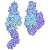
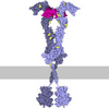
- PDB-11yb: Structure of AlphaIIbBeta3-R21C11 Fab in the near-bent conformation -
+ Open data

- Basic information
| Entry | Database: PDB / ID: 11yb | |||||||||
|---|---|---|---|---|---|---|---|---|---|---|
| Title | Structure of AlphaIIbBeta3-R21C11 Fab in the near-bent conformation | |||||||||
 Components Components |
| |||||||||
 Keywords Keywords | BLOOD CLOTTING / Monoclonal antibody / integrin receptor / platelets / inhibiter of PDI binding to AlphaIIbbeta3 integrin | |||||||||
| Function / homology |  Function and homology information Function and homology informationregulation of serotonin uptake / positive regulation of adenylate cyclase-inhibiting opioid receptor signaling pathway / tube development / alpha9-beta1 integrin-ADAM8 complex / response to platelet-derived growth factor / integrin alphaIIb-beta3 complex / regulation of postsynaptic neurotransmitter receptor diffusion trapping / maintenance of postsynaptic specialization structure / alphav-beta3 integrin-vitronectin complex / platelet alpha granule membrane ...regulation of serotonin uptake / positive regulation of adenylate cyclase-inhibiting opioid receptor signaling pathway / tube development / alpha9-beta1 integrin-ADAM8 complex / response to platelet-derived growth factor / integrin alphaIIb-beta3 complex / regulation of postsynaptic neurotransmitter receptor diffusion trapping / maintenance of postsynaptic specialization structure / alphav-beta3 integrin-vitronectin complex / platelet alpha granule membrane / integrin alphav-beta3 complex / negative regulation of lipoprotein metabolic process / positive regulation of glomerular mesangial cell proliferation / smooth muscle cell migration / alphav-beta3 integrin-PKCalpha complex / fibrinogen binding / positive regulation of leukocyte migration / alphav-beta3 integrin-HMGB1 complex / negative regulation of lipid transport / vascular endothelial growth factor receptor 2 binding / angiogenesis involved in wound healing / positive regulation of vascular endothelial growth factor signaling pathway / regulation of release of sequestered calcium ion into cytosol / Elastic fibre formation / mesodermal cell differentiation / positive regulation of bone resorption / alphav-beta3 integrin-IGF-1-IGF1R complex / platelet-derived growth factor receptor binding / cell-cell adhesion mediated by integrin / filopodium membrane / glycinergic synapse / extracellular matrix binding / positive regulation of fibroblast migration / positive regulation of cell adhesion mediated by integrin / positive regulation of vascular endothelial growth factor receptor signaling pathway / apolipoprotein A-I-mediated signaling pathway / regulation of bone resorption / negative regulation of low-density lipoprotein particle clearance / apoptotic cell clearance / wound healing, spreading of epidermal cells / positive regulation of smooth muscle cell migration / integrin complex / Molecules associated with elastic fibres / heterotypic cell-cell adhesion / cell adhesion mediated by integrin / negative chemotaxis / positive regulation of osteoblast proliferation / Mechanical load activates signaling by PIEZO1 and integrins in osteocytes / cellular response to insulin-like growth factor stimulus / Syndecan interactions / p130Cas linkage to MAPK signaling for integrins / regulation of postsynaptic neurotransmitter receptor internalization / cell-substrate adhesion / protein disulfide isomerase activity / microvillus membrane / PECAM1 interactions / GRB2:SOS provides linkage to MAPK signaling for Integrins / negative regulation of endothelial cell apoptotic process / TGF-beta receptor signaling activates SMADs / fibronectin binding / lamellipodium membrane / negative regulation of macrophage derived foam cell differentiation / negative regulation of lipid storage / Integrin cell surface interactions / ECM proteoglycans / substrate adhesion-dependent cell spreading / positive regulation of T cell migration / cell-matrix adhesion / embryo implantation / coreceptor activity / Integrin signaling / positive regulation of endothelial cell proliferation / positive regulation of smooth muscle cell proliferation / positive regulation of endothelial cell migration / cell adhesion molecule binding / integrin-mediated signaling pathway / Turbulent (oscillatory, disturbed) flow shear stress activates signaling by PIEZO1 and integrins in endothelial cells / response to activity / protein kinase C binding / wound healing / Signal transduction by L1 / cellular response to xenobiotic stimulus / regulation of actin cytoskeleton organization / cell-cell adhesion / cellular response to mechanical stimulus / platelet activation / Signaling by high-kinase activity BRAF mutants / RUNX1 regulates genes involved in megakaryocyte differentiation and platelet function / MAP2K and MAPK activation / platelet aggregation / VEGFA-VEGFR2 Pathway / integrin binding / ruffle membrane / blood coagulation / positive regulation of angiogenesis / Signaling by RAF1 mutants / Signaling by moderate kinase activity BRAF mutants / Paradoxical activation of RAF signaling by kinase inactive BRAF / Signaling downstream of RAS mutants / cell-cell junction Similarity search - Function | |||||||||
| Biological species |  Homo sapiens (human) Homo sapiens (human)  Mus musculus (house mouse) Mus musculus (house mouse) | |||||||||
| Method | ELECTRON MICROSCOPY / single particle reconstruction / cryo EM / Resolution: 3.4 Å | |||||||||
 Authors Authors | Wang, J.L. / Coller, B. / Wang, L. / Li, J.H. | |||||||||
| Funding support |  United States, 1items United States, 1items
| |||||||||
 Citation Citation | Journal: Blood Adv / Year: 2026 Title: A monoclonal antibody to platelet αIIbβ3 that inhibits protein disulfide isomerase binding and platelet aggregation. Authors: Lu Wang / Jialing Wang / Jihong Li / Barry S Coller / Abstract: Platelet integrin αIIbβ3 plays a pivotal role in hemostasis and thrombosis. Protein disulfide isomerase (PDI) binds to αIIbβ3 and mediates its activation. The binding region(s) on αIIbβ3 for ...Platelet integrin αIIbβ3 plays a pivotal role in hemostasis and thrombosis. Protein disulfide isomerase (PDI) binds to αIIbβ3 and mediates its activation. The binding region(s) on αIIbβ3 for PDI remains to be established. We identified a new anti-αIIbβ3 murine monoclonal antibody, R21C11, that inhibits PDI binding to platelets, ligand binding to αIIbβ3, and platelet aggregation. R21C11 inhibited recombinant PDI binding to platelets activated with the PAR1 thrombin receptor activating peptide SFLLRN (T6). Reciprocally, PDI partially reduced R21C11 binding to platelets. PDI was translocated to the platelet surface after activation and activated platelets showed higher PDI reductase activity. R211C11 decreased both the amount of PDI and the reductase activity. R21C11 partially inhibited both fibrinogen and PAC-1 binding to αIIbβ3 and platelet aggregation induced by ADP and T6. R21C11 bound slowly to unactivated platelets and more rapidly after T6 activation; eptifibatide, which induces the αIIbβ3 extended-open conformation, also increased the speed of R21C11 binding. Activation also increased total R21C11 binding. Cryogenic electron microscopy single-particle analysis of the R21C11 Fab-αIIbβ3 complex revealed that R21C11 binds to the β3 β-tail domain in both nearly bent and semiextended-closed conformations of αIIbβ3. R21C11 Fab clashed with the β3 β-I domain in the fully bent conformation, accounting for the slow binding rate of R21C11. These data are consistent with R21C11 inhibiting PDI binding by steric hindrance and the activation dependence of PDI binding. Taken together, these data suggest that the β-tail domain and/or neighboring area is the binding site for PDI on integrin αIIbβ3. | |||||||||
| History |
|
- Structure visualization
| Structure viewer | Molecule: MolmilJmol/JSmol |
|---|
- Downloads & links
-Download
| PDBx/mmCIF format | 11yb.cif.gz | 558.1 KB | Display | PDBx/mmCIF format |
|---|---|---|---|---|
| PDB format | pdb11yb.ent.gz | 448.9 KB | Display | PDB format |
| PDBx/mmJSON format | 11yb.json.gz | Tree view | PDBx/mmJSON format | |
| Others |  Other downloads Other downloads |
-Validation report
| Arichive directory | https://data.pdbj.org/pub/pdb/validation_reports/1y/11ybftp://data.pdbj.org/pub/pdb/validation_reports/1y/11yb | HTTPS FTP |
|---|
-Related structure data
| Related structure data |  76179MC M: map data used to model this data C: citing same article ( |
|---|---|
| Similar structure data |
-Links
 PDBj
PDBj









- Assembly
| Deposited unit | 
|
|---|---|
| 1 |
|
-Components
-Protein , 2 types, 2 molecules AB
| #1: Protein | Mass: 113477.523 Da / Num. of mol.: 1 Source method: isolated from a genetically manipulated source Source: (gene. exp.) Homo sapiens (human) / Gene: ITGA2B, GP2B, ITGAB / Production host: Homo sapiens (human) / References: UniProt: P08514 |
|---|---|
| #2: Protein | Mass: 87150.773 Da / Num. of mol.: 1 Source method: isolated from a genetically manipulated source Source: (gene. exp.) Homo sapiens (human) / Gene: ITGB3, GP3A / Production host: Homo sapiens (human) / References: UniProt: P05106 |
-Antibody , 2 types, 2 molecules HL
| #3: Antibody | Mass: 23931.814 Da / Num. of mol.: 1 Source method: isolated from a genetically manipulated source Source: (gene. exp.) Mus musculus (house mouse) / Production host: Mus musculus (house mouse) |
|---|---|
| #4: Antibody | Mass: 23218.652 Da / Num. of mol.: 1 Source method: isolated from a genetically manipulated source Details: LLTQSPATLSVTPGETVSLSCRASQSIYKNLHWYQQKSHRSPSLLIKSASESISGIPSRFTGSGSGTDYILSINSVEPEDEGIYYCLQGYSIPFTFGAGSKLELKRADAAPTVSIFPPSSEQLTSGGASVVCFLNNFYPKDLNVKWKLDGSERQNGVLDSWTDQDSKDSTYMSSTLTLTKDEYERHNSYTCEATHKTSTSPLVKSFNRNEC Source: (gene. exp.) Mus musculus (house mouse) / Production host: Mus musculus (house mouse) |
-Non-polymers , 2 types, 8 molecules 

| #5: Chemical | ChemComp-CA /  Mass: 40.078 Da / Num. of mol.: 7 / Source method: obtained synthetically / Formula: Ca Mass: 40.078 Da / Num. of mol.: 7 / Source method: obtained synthetically / Formula: Ca#6: Chemical | ChemComp-MG / |  Mass: 24.305 Da / Num. of mol.: 1 / Source method: obtained synthetically / Formula: Mg Mass: 24.305 Da / Num. of mol.: 1 / Source method: obtained synthetically / Formula: Mg |
|---|
-Details
| Has ligand of interest | N |
|---|---|
| Has protein modification | Y |
-Experimental details
-Experiment
| Experiment | Method: ELECTRON MICROSCOPY |
|---|---|
| EM experiment | Aggregation state: PARTICLE / 3D reconstruction method: single particle reconstruction |
- Sample preparation
| Component |
| ||||||||||||||||||||||||
|---|---|---|---|---|---|---|---|---|---|---|---|---|---|---|---|---|---|---|---|---|---|---|---|---|---|
| Molecular weight |
| ||||||||||||||||||||||||
| Source (natural) |
| ||||||||||||||||||||||||
| Buffer solution | pH: 7.4 | ||||||||||||||||||||||||
| Specimen | Conc.: 0.05 mg/ml / Embedding applied: NO / Shadowing applied: NO / Staining applied: NO / Vitrification applied: YES | ||||||||||||||||||||||||
| Specimen support | Grid material: COPPER / Grid mesh size: 400 divisions/in. / Grid type: Quantifoil R1.2/1.3 | ||||||||||||||||||||||||
| Vitrification | Instrument: FEI VITROBOT MARK I / Cryogen name: ETHANE / Humidity: 100 % / Chamber temperature: 277 K |
- Electron microscopy imaging
| Experimental equipment |  Model: Titan Krios / Image courtesy: FEI Company |
|---|---|
| Microscopy | Model: TFS KRIOS |
| Electron gun | Electron source:  FIELD EMISSION GUN / Accelerating voltage: 300 kV / Illumination mode: FLOOD BEAM FIELD EMISSION GUN / Accelerating voltage: 300 kV / Illumination mode: FLOOD BEAM |
| Electron lens | Mode: BRIGHT FIELD / Nominal magnification: 81000 X / Nominal defocus max: 2500 nm / Nominal defocus min: 1500 nm / Alignment procedure: COMA FREE |
| Specimen holder | Cryogen: NITROGEN / Specimen holder model: FEI TITAN KRIOS AUTOGRID HOLDER |
| Image recording | Electron dose: 54 e/Å2 / Film or detector model: GATAN K3 (6k x 4k) |
- Processing
| EM software |
| ||||||||||||||||||||||||||||||||||||
|---|---|---|---|---|---|---|---|---|---|---|---|---|---|---|---|---|---|---|---|---|---|---|---|---|---|---|---|---|---|---|---|---|---|---|---|---|---|
| CTF correction | Type: PHASE FLIPPING AND AMPLITUDE CORRECTION | ||||||||||||||||||||||||||||||||||||
| Particle selection | Num. of particles selected: 1332000 | ||||||||||||||||||||||||||||||||||||
| 3D reconstruction | Resolution: 3.4 Å / Resolution method: FSC 0.143 CUT-OFF / Num. of particles: 49648 / Algorithm: FOURIER SPACE / Num. of class averages: 1 / Symmetry type: POINT | ||||||||||||||||||||||||||||||||||||
| Atomic model building | Protocol: RIGID BODY FIT / Space: REAL | ||||||||||||||||||||||||||||||||||||
| Atomic model building | 3D fitting-ID: 1 / Source name: PDB / Type: experimental model
|


