 Movie
Movie Controller


+ Open data

- Basic information
| Entry | Database: PDB / ID: 11vg | ||||||||||||
|---|---|---|---|---|---|---|---|---|---|---|---|---|---|
| Title | D189F thrombin inhibited with D-Phe-Pro-Gln-Chloromethylketone | ||||||||||||
 Components Components |
| ||||||||||||
 Keywords Keywords | BLOOD CLOTTING / serine protease / inhibitor | ||||||||||||
| Function / homology |  Function and homology information Function and homology informationnegative regulation of astrocyte differentiation / thrombospondin receptor activity / thrombin / thrombin-activated receptor signaling pathway / Defective factor XII causes hereditary angioedema / regulation of blood coagulation / neutrophil-mediated killing of gram-negative bacterium / positive regulation of phospholipase C-activating G protein-coupled receptor signaling pathway / Platelet Aggregation (Plug Formation) / Defective F8 cleavage by thrombin ...negative regulation of astrocyte differentiation / thrombospondin receptor activity / thrombin / thrombin-activated receptor signaling pathway / Defective factor XII causes hereditary angioedema / regulation of blood coagulation / neutrophil-mediated killing of gram-negative bacterium / positive regulation of phospholipase C-activating G protein-coupled receptor signaling pathway / Platelet Aggregation (Plug Formation) / Defective F8 cleavage by thrombin / positive regulation of collagen biosynthetic process / Fibrin formation / negative regulation of platelet activation / negative regulation of blood coagulation / negative regulation of fibrinolysis / positive regulation of blood coagulation / negative regulation of proteolysis / Transport of gamma-carboxylated protein precursors from the endoplasmic reticulum to the Golgi apparatus / fibrinolysis / Gamma-carboxylation of protein precursors / Amplification and propagation of coagulation cascade / Removal of aminoterminal propeptides from gamma-carboxylated proteins / Initiation of coagulation cascade / regulation of cytosolic calcium ion concentration / negative regulation of cytokine production involved in inflammatory response / Regulation of clotting cascade / Regulation of Complement cascade / positive regulation of receptor signaling pathway via JAK-STAT / positive regulation of release of sequestered calcium ion into cytosol / Cell surface interactions at the vascular wall / growth factor activity / Peptide ligand-binding receptors / response to wounding / lipopolysaccharide binding / platelet activation / positive regulation of protein localization to nucleus / Golgi lumen / blood coagulation / Regulation of Insulin-like Growth Factor (IGF) transport and uptake by Insulin-like Growth Factor Binding Proteins (IGFBPs) / positive regulation of reactive oxygen species metabolic process / heparin binding / Thrombin signalling through proteinase activated receptors (PARs) / Dengue Virus-Host Interactions / antimicrobial humoral immune response mediated by antimicrobial peptide / blood microparticle / extracellular matrix / G alpha (q) signalling events / cell surface receptor signaling pathway / receptor ligand activity / endoplasmic reticulum lumen / serine-type endopeptidase activity / signaling receptor binding / calcium ion binding / proteolysis / extracellular exosome / extracellular region / plasma membrane Similarity search - Function | ||||||||||||
| Biological species |  Homo sapiens (human) Homo sapiens (human) | ||||||||||||
| Method |  X-RAY DIFFRACTION / SYNCHROTRON / MOLECULAR REPLACEMENT / Resolution: 2.13 Å X-RAY DIFFRACTION / SYNCHROTRON / MOLECULAR REPLACEMENT / Resolution: 2.13 Å | ||||||||||||
 Authors Authors | Friet, T. / Mohammed, B.M. / Sukumar, N. / Di Cera, E. | ||||||||||||
| Funding support |  United States, 3items United States, 3items
| ||||||||||||
 Citation Citation | Journal: J.Thromb.Haemost. / Year: 2026 Title: Structural analysis of the primary specificity of thrombin. Authors: Friet, T. / Mikhail, G. / Mohammed, B.M. / Pelc, L.A. / Dei Rossi, A. / Korolev, S. / Di Cera, E. | ||||||||||||
| History |
|
- Structure visualization
| Structure viewer | Molecule: MolmilJmol/JSmol |
|---|
- Downloads & links
-Download
| PDBx/mmCIF format | 11vg.cif.gz | 308 KB | Display | PDBx/mmCIF format |
|---|---|---|---|---|
| PDB format | pdb11vg.ent.gz | Display | PDB format | |
| PDBx/mmJSON format | 11vg.json.gz | Tree view | PDBx/mmJSON format | |
| Others |  Other downloads Other downloads |
-Validation report
| Arichive directory | https://data.pdbj.org/pub/pdb/validation_reports/1v/11vgftp://data.pdbj.org/pub/pdb/validation_reports/1v/11vg | HTTPS FTP |
|---|
-Related structure data
| Related structure data |  11riC  11rjC  11rkC  11xwC  11yhC  12cwC  12cxC  12cyC  12itC C: citing same article ( |
|---|---|
| Similar structure data |
-Links
 PDBj
PDBj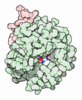



- Assembly
| Deposited unit | 
| ||||||||||||
|---|---|---|---|---|---|---|---|---|---|---|---|---|---|
| 1 | 
| ||||||||||||
| 2 | 
| ||||||||||||
| Unit cell |
|
-Components
| #1: Protein/peptide | Mass: 5641.175 Da / Num. of mol.: 2 Source method: isolated from a genetically manipulated source Source: (gene. exp.) Homo sapiens (human) / Gene: F2 / Production host:  Mesocricetus auratus (golden hamster) / References: UniProt: P00734 Mesocricetus auratus (golden hamster) / References: UniProt: P00734#2: Protein | Mass: 31457.119 Da / Num. of mol.: 2 / Mutation: D519F Source method: isolated from a genetically manipulated source Source: (gene. exp.) Homo sapiens (human) / Gene: F2 / Production host: Mesocricetus auratus (golden hamster) / References: UniProt: P00734#3: Sugar |   Type: D-saccharide, beta linking / Mass: 221.208 Da / Num. of mol.: 2 Type: D-saccharide, beta linking / Mass: 221.208 Da / Num. of mol.: 2Source method: isolated from a genetically manipulated source Formula: C8H15NO6 #4: Chemical | Mass: 424.922 Da / Num. of mol.: 2 / Source method: obtained synthetically / Formula: C20H29ClN4O4 / Feature type: SUBJECT OF INVESTIGATION #5: Water | ChemComp-HOH / |  Mass: 18.015 Da / Num. of mol.: 279 / Source method: isolated from a natural source / Formula: H2O Mass: 18.015 Da / Num. of mol.: 279 / Source method: isolated from a natural source / Formula: H2OHas ligand of interest | Y | Has protein modification | Y | |
|---|
-Experimental details
-Experiment
| Experiment | Method: X-RAY DIFFRACTION / Number of used crystals: 1 |
|---|
- Sample preparation
| Crystal | Density Matthews: 2.61 Å3/Da / Density % sol: 52.86 % |
|---|---|
| Crystal grow | Temperature: 293.15 K / Method: vapor diffusion, hanging drop Details: 0.1 M BIS-TRIS pH 6.5, 20% w/v Polyethylene glycol monomethyl ether 5,000 |
-Data collection
| Diffraction | Mean temperature: 100 K / Serial crystal experiment: N |
|---|---|
| Diffraction source | Source: SYNCHROTRON / Site: NSLS-II / Beamline: 17-ID-2 / Wavelength: 0.97934 Å |
| Detector | Type: DECTRIS EIGER X 16M / Detector: PIXEL / Date: Jul 15, 2025 |
| Radiation | Protocol: SINGLE WAVELENGTH / Monochromatic (M) / Laue (L): M / Scattering type: x-ray |
| Radiation wavelength | Wavelength: 0.97934 Å / Relative weight: 1 |
| Reflection | Resolution: 2.13→33.65 Å / Num. obs: 40358 / % possible obs: 98.3 % / Redundancy: 6 % / Biso Wilson estimate: 10.98 Å2 / CC1/2: 0.93 / Rmerge(I) obs: 0.43 / Rpim(I) all: 0.186 / Rrim(I) all: 0.47 / Χ2: 0.89 / Net I/σ(I): 3.2 / Num. measured all: 240634 |
| Reflection shell | Resolution: 2.13→2.25 Å / % possible obs: 97.3 % / Redundancy: 5.8 % / Rmerge(I) obs: 1.395 / Num. measured all: 33790 / Num. unique obs: 5855 / CC1/2: 0.374 / Rpim(I) all: 0.604 / Rrim(I) all: 1.523 / Χ2: 0.84 / Net I/σ(I) obs: 1.5 |
- Processing
| Software |
| |||||||||||||||||||||||||||||||||||||||||||||||||||||||||||||||||||||||||||||||||||||||||||||||||||||||||
|---|---|---|---|---|---|---|---|---|---|---|---|---|---|---|---|---|---|---|---|---|---|---|---|---|---|---|---|---|---|---|---|---|---|---|---|---|---|---|---|---|---|---|---|---|---|---|---|---|---|---|---|---|---|---|---|---|---|---|---|---|---|---|---|---|---|---|---|---|---|---|---|---|---|---|---|---|---|---|---|---|---|---|---|---|---|---|---|---|---|---|---|---|---|---|---|---|---|---|---|---|---|---|---|---|---|---|
| Refinement | Method to determine structure: MOLECULAR REPLACEMENT / Resolution: 2.13→33.65 Å / SU ML: 0.2848 / Cross valid method: FREE R-VALUE / σ(F): 1.39 / Phase error: 24.7955 Stereochemistry target values: GeoStd + Monomer Library + CDL v1.2
| |||||||||||||||||||||||||||||||||||||||||||||||||||||||||||||||||||||||||||||||||||||||||||||||||||||||||
| Solvent computation | Shrinkage radii: 0.9 Å / VDW probe radii: 1.1 Å / Solvent model: FLAT BULK SOLVENT MODEL | |||||||||||||||||||||||||||||||||||||||||||||||||||||||||||||||||||||||||||||||||||||||||||||||||||||||||
| Displacement parameters | Biso mean: 14.21 Å2 | |||||||||||||||||||||||||||||||||||||||||||||||||||||||||||||||||||||||||||||||||||||||||||||||||||||||||
| Refinement step | Cycle: LAST / Resolution: 2.13→33.65 Å
| |||||||||||||||||||||||||||||||||||||||||||||||||||||||||||||||||||||||||||||||||||||||||||||||||||||||||
| Refine LS restraints |
| |||||||||||||||||||||||||||||||||||||||||||||||||||||||||||||||||||||||||||||||||||||||||||||||||||||||||
| LS refinement shell |
| |||||||||||||||||||||||||||||||||||||||||||||||||||||||||||||||||||||||||||||||||||||||||||||||||||||||||
| Refinement TLS params. | Method: refined / Origin x: 16.322406169684 Å / Origin y: 40.371359452131 Å / Origin z: 0.03121201372925 Å
| |||||||||||||||||||||||||||||||||||||||||||||||||||||||||||||||||||||||||||||||||||||||||||||||||||||||||
| Refinement TLS group | Selection details: all |
