[English] 日本語
Yorodumi
- PDB-10fv: Crystal Structure of Human Fgr SH3-SH2-High Affinity Linker Mutan... -

+
Open data


ID or keywords:

Loading...

-
Basic information

Entry
Database: PDB / ID: 10fv
TitleCrystal Structure of Human Fgr SH3-SH2-High Affinity Linker Mutant 2 (HAL2) Domains.
ComponentsTyrosine-protein kinase Fgr
KeywordsTRANSFERASE / FGR / SIGNALING PROTEIN / SH3 / SH2
Function / homology
Function and homology information


negative regulation of natural killer cell activation / positive regulation of mast cell degranulation / immune response-regulating cell surface receptor signaling pathway / regulation of protein kinase activity / regulation of phagocytosis / Fc-gamma receptor I complex binding / immunoglobulin receptor binding / Platelet sensitization by LDL / regulation of innate immune response / Fc-gamma receptor signaling pathway involved in phagocytosis ...negative regulation of natural killer cell activation / positive regulation of mast cell degranulation / immune response-regulating cell surface receptor signaling pathway / regulation of protein kinase activity / regulation of phagocytosis / Fc-gamma receptor I complex binding / immunoglobulin receptor binding / Platelet sensitization by LDL / regulation of innate immune response / Fc-gamma receptor signaling pathway involved in phagocytosis / FCGR activation / phosphotyrosine residue binding / peptidyl-tyrosine phosphorylation / FCGR3A-mediated IL10 synthesis / cell surface receptor protein tyrosine kinase signaling pathway / integrin-mediated signaling pathway / positive regulation of cytokine production / non-specific protein-tyrosine kinase / FCGR3A-mediated phagocytosis / negative regulation of inflammatory response to antigenic stimulus / non-membrane spanning protein tyrosine kinase activity / mitochondrial intermembrane space / ruffle membrane / response to virus / protein autophosphorylation / regulation of cell shape / actin cytoskeleton / protein tyrosine kinase activity / secretory granule lumen / protein phosphorylation / cell differentiation / positive regulation of phosphatidylinositol 3-kinase/protein kinase B signal transduction / mitochondrial inner membrane / defense response to Gram-positive bacterium / positive regulation of cell migration / signaling receptor binding / innate immune response / Neutrophil degranulation / protein kinase binding / extracellular exosome / extracellular region / ATP binding / plasma membrane / cytosol
Similarity search - Function
Tyrosine-protein kinase Fgr, SH2 domain / : / SH3 domain / SH2 domain / Src homology 2 (SH2) domain profile. / Src homology 2 domains / SH2 domain / Src homology 3 domains / SH2 domain superfamily / SH3-like domain superfamily ...Tyrosine-protein kinase Fgr, SH2 domain / : / SH3 domain / SH2 domain / Src homology 2 (SH2) domain profile. / Src homology 2 domains / SH2 domain / Src homology 3 domains / SH2 domain superfamily / SH3-like domain superfamily / Src homology 3 (SH3) domain profile. / SH3 domain / Tyrosine-protein kinase, catalytic domain / Tyrosine kinase, catalytic domain / Tyrosine protein kinases specific active-site signature. / Tyrosine-protein kinase, active site / Protein tyrosine and serine/threonine kinase / Serine-threonine/tyrosine-protein kinase, catalytic domain / Protein kinase, ATP binding site / Protein kinases ATP-binding region signature. / Protein kinase domain profile. / Protein kinase domain / Protein kinase-like domain superfamily
Similarity search - Domain/homology
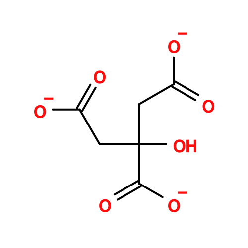
CITRATE ANION / Tyrosine-protein kinase Fgr
Similarity search - Component
Biological speciesHomo sapiens (human)
MethodX-RAY DIFFRACTION / SYNCHROTRON / MOLECULAR REPLACEMENT / Resolution: 1.8 Å
AuthorsAlvarado, J.J. / Gonzalez-Areizaga, G. / Smithgall, T.E.
Funding support United States, 2items
OrganizationGrant numberCountry
National Institutes of Health/National Cancer Institute (NIH/NCI)CA233576 United States
National Institutes of Health/National Cancer Institute (NIH/NCI)CA291724 United States
CitationJournal: Cell Rep / Year: 2026
Title: Constraining regulatory domain dynamics of the Src kinase Fgr increases ATP-site inhibitor sensitivity and impairs bone marrow engraftment.
Authors: Gonzalez-Areizaga, G. / Shu, S.T. / Alvarado, J.J. / Shi, H. / Chen, L. / Smithgall, T.E.
History
DepositionJan 18, 2026Deposition site: RCSB / Processing site: RCSB
Revision 1.0Jul 15, 2026Provider: repository / Type: Initial release

-
Structure visualization

Structure viewerMolecule:
MolmilJmol/JSmol

Downloads & links

-
Assembly

Deposited unit
A: Tyrosine-protein kinase Fgr
hetero molecules


Theoretical massNumber of molelcules
Total (without water)21,51713
Polymers20,3071
Non-polymers1,21012
Water61334
1


  • Idetical with deposited unit
  • defined by author&software
  • Evidence: gel filtration
TypeNameSymmetry operationNumber
identity operation1_555x,y,z1
MethodPISA
Unit cell
Length a, b, c (Å)43.393, 35.127, 60.846
Angle α, β, γ (deg.)90, 100.642, 90
Int Tables number4
Space group name H-MP1211

-
Components

#1: Protein Tyrosine-protein kinase Fgr / Gardner-Rasheed feline sarcoma viral (v-fgr) oncogene homolog / Proto-oncogene c-Fgr / p55-Fgr / ...Gardner-Rasheed feline sarcoma viral (v-fgr) oncogene homolog / Proto-oncogene c-Fgr / p55-Fgr / p58-Fgr / p58c-Fgr


Mass: 20306.855 Da / Num. of mol.: 1 / Mutation: A256R
Source method: isolated from a genetically manipulated source
Details: Human Fgr SH3, SH2 and linker domains; the wild-type linker residue A256 was mutated to R256. N-terminal serine residue originates from Sumo tag Ulp1 cleavage. Residue C131 was covalently ...Details: Human Fgr SH3, SH2 and linker domains; the wild-type linker residue A256 was mutated to R256. N-terminal serine residue originates from Sumo tag Ulp1 cleavage. Residue C131 was covalently modified with beta mercaptoethanol reducing agent during the purification process.
Source: (gene. exp.) Homo sapiens (human) / Gene: FGR, SRC2 / Production host: Escherichia coli (E. coli) / Variant (production host): BL21(DE3) STAR
References: UniProt: P09769, non-specific protein-tyrosine kinase
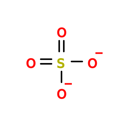
#2: Chemical ChemComp-SO4 / SULFATE ION


Mass: 96.063 Da / Num. of mol.: 2 / Source method: obtained synthetically / Formula: SO4
#3: Chemical
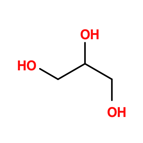
ChemComp-GOL / GLYCEROL / GLYCERIN / PROPANE-1,2,3-TRIOL


Mass: 92.094 Da / Num. of mol.: 9 / Source method: obtained synthetically / Formula: C3H8O3
#4: Chemical ChemComp-FLC / CITRATE ANION


Mass: 189.100 Da / Num. of mol.: 1 / Source method: obtained synthetically / Formula: C6H5O7
#5: Water ChemComp-HOH / water


Mass: 18.015 Da / Num. of mol.: 34 / Source method: isolated from a natural source / Formula: H2O
Has ligand of interestN
Has protein modificationY

-
Experimental details

-
Experiment

ExperimentMethod: X-RAY DIFFRACTION / Number of used crystals: 1

-
Sample preparation

CrystalDensity Matthews: 2.26 Å3/Da / Density % sol: 45.5 %
Crystal growTemperature: 298.15 K / Method: vapor diffusion, sitting drop / pH: 5.5
Details: 0.1 M trisodium citrate pH 5.5, 2.5 M ammonium sulfate

-
Data collection

DiffractionMean temperature: 100 K / Serial crystal experiment: N
Diffraction sourceSource: SYNCHROTRON / Site: APS / Beamline: 23-ID-D / Wavelength: 1 Å
DetectorType: DECTRIS EIGER2 X CdTe 16M / Detector: PIXEL / Date: Jul 28, 2021 / Details: kb bent focusing mirror
RadiationMonochromator: double crystal monochromator / Protocol: SINGLE WAVELENGTH / Monochromatic (M) / Laue (L): M / Scattering type: x-ray
Radiation wavelengthWavelength: 1 Å / Relative weight: 1
ReflectionResolution: 1.8→42.65 Å / Num. obs: 16998 / % possible obs: 100 % / Redundancy: 1.9 % / CC1/2: 0.999 / Rmerge(I) obs: 0.029 / Rpim(I) all: 0.029 / Rrim(I) all: 0.041 / Χ2: 0.58 / Net I/σ(I): 9.7
Reflection shell

Diffraction-ID: 1

Resolution (Å)Redundancy (%)Rmerge(I) obsMean I/σ(I) obsNum. unique obsCC1/2Rpim(I) allRrim(I) allΧ2% possible all
9-42.651.70.01128.71550.9990.0110.0160.3399.2
1.8-1.8420.6211.19990.4850.6210.8790.67100

-
Processing

Software
NameVersionClassification
REFMAC5.8.0431 (refmacat 0.4.126)refinement
Cootmodel building
PHASERphasing
Aimlessdata scaling
xia2data reduction
RefinementMethod to determine structure: MOLECULAR REPLACEMENT / Resolution: 1.8→42.65 Å / Cor.coef. Fo:Fc: 0.966 / Cor.coef. Fo:Fc free: 0.95 / SU B: 7.835 / SU ML: 0.109 / Cross valid method: FREE R-VALUE / ESU R: 0.127 / ESU R Free: 0.124
Details: Hydrogens have been added in their riding positions
RfactorNum. reflection% reflectionSelection details
Rfree0.2331 883 5.198 %RANDOM
Rwork0.1944 16104 --
all0.196 ---
obs-16987 99.959 %-
Solvent computationIon probe radii: 0.8 Å / Shrinkage radii: 0.8 Å / VDW probe radii: 1.2 Å / Solvent model: MASK BULK SOLVENT
Displacement parametersBiso mean: 45.797 Å2
Baniso -1Baniso -2Baniso -3
1--1.24 Å20 Å2-0.39 Å2
2---1.105 Å20 Å2
3---2.327 Å2
Refinement stepCycle: LAST / Resolution: 1.8→42.65 Å
ProteinNucleic acidLigandSolventTotal
Num. atoms1251 0 77 34 1362
Refine LS restraints
Refine-IDTypeDev idealDev ideal targetNumber
X-RAY DIFFRACTIONr_bond_refined_d0.0080.0121346
X-RAY DIFFRACTIONr_bond_other_d0.0010.0161212
X-RAY DIFFRACTIONr_angle_refined_deg1.6291.811807
X-RAY DIFFRACTIONr_angle_other_deg0.5711.7642808
X-RAY DIFFRACTIONr_dihedral_angle_1_deg7.4965153
X-RAY DIFFRACTIONr_dihedral_angle_2_deg9.06358
X-RAY DIFFRACTIONr_dihedral_angle_3_deg14.51610212
X-RAY DIFFRACTIONr_dihedral_angle_6_deg12.61064
X-RAY DIFFRACTIONr_chiral_restr0.080.2195
X-RAY DIFFRACTIONr_gen_planes_refined0.0070.021534
X-RAY DIFFRACTIONr_gen_planes_other0.0010.02310
X-RAY DIFFRACTIONr_nbd_refined0.1950.2209
X-RAY DIFFRACTIONr_symmetry_nbd_other0.2010.21044
X-RAY DIFFRACTIONr_nbtor_refined0.1920.2607
X-RAY DIFFRACTIONr_symmetry_nbtor_other0.0870.2727
X-RAY DIFFRACTIONr_xyhbond_nbd_refined0.1160.233
X-RAY DIFFRACTIONr_symmetry_nbd_refined0.2320.210
X-RAY DIFFRACTIONr_nbd_other0.2190.248
X-RAY DIFFRACTIONr_symmetry_xyhbond_nbd_refined0.1630.24
X-RAY DIFFRACTIONr_mcbond_it2.1642.464618
X-RAY DIFFRACTIONr_mcbond_other2.1512.465618
X-RAY DIFFRACTIONr_mcangle_it3.1034.394769
X-RAY DIFFRACTIONr_mcangle_other3.1024.402770
X-RAY DIFFRACTIONr_scbond_it3.4193.111728
X-RAY DIFFRACTIONr_scbond_other3.4173.117729
X-RAY DIFFRACTIONr_scangle_it4.9685.4721038
X-RAY DIFFRACTIONr_scangle_other4.9655.4781039
X-RAY DIFFRACTIONr_lrange_it7.37327.8211367
X-RAY DIFFRACTIONr_lrange_other7.40927.4251365
LS refinement shell

Refine-ID: X-RAY DIFFRACTION / Total num. of bins used: 20

Resolution (Å)Rfactor RfreeNum. reflection RfreeRfactor RworkNum. reflection RworkRfactor allNum. reflection allFsc freeFsc work% reflection obs (%)WRfactor Rwork
1.8-1.8470.388660.35311950.35512610.9120.9121000.351
1.847-1.8970.429640.34311260.34711900.9030.9231000.34
1.897-1.9520.368610.30911270.31211900.9120.94199.83190.303
1.952-2.0120.285570.26910780.2711350.930.9541000.26
2.012-2.0780.234560.22310610.22411170.9660.9691000.214
2.078-2.1510.267540.21210050.21510590.9570.9711000.201
2.151-2.2320.293420.20410080.20810510.9530.97499.90490.193
2.232-2.3220.203370.1879690.18710070.9830.97999.90070.175
2.322-2.4250.239540.1918970.1949510.9650.9791000.178
2.425-2.5430.239530.1818780.1859310.9770.981000.171
2.543-2.680.261480.2068180.2098660.9640.9741000.198
2.68-2.8420.299470.2127780.2178250.9490.9741000.204
2.842-3.0380.246410.2027360.2047770.9660.9741000.199
3.038-3.280.224440.1846940.1867390.970.9899.86470.191
3.28-3.5910.204360.1716470.1736830.9750.9821000.189
3.591-4.0110.194350.1635710.1646060.9810.9841000.185
4.011-4.6240.216270.1425180.1455460.9750.98899.81680.164
4.624-5.6470.203280.184390.1824670.9750.9841000.22
5.647-7.9170.191200.213480.2093680.9770.9731000.24
7.917-42.650.187130.2172110.2162240.9740.9691000.302
Refinement TLS params.

Method: refined / Refine-ID: X-RAY DIFFRACTION

IDL112)L122)L132)L222)L232)L332)S11 (Å °)S12 (Å °)S13 (Å °)S21 (Å °)S22 (Å °)S23 (Å °)S31 (Å °)S32 (Å °)S33 (Å °)T112)T122)T132)T222)T232)T332)Origin x (Å)Origin y (Å)Origin z (Å)
14.0774-0.86610.4553.85790.85884.2304-0.0040.13520.124-0.0332-0.0256-0.0723-0.05850.18260.02950.0598-0.00370.00750.03610.00440.00550.56592.500530.3821
24.9398-2.121.60245.0729-0.99651.7530.170.33170.0039-0.3177-0.17350.28430.0047-0.14190.00360.2559-0.0038-0.01230.3305-0.03960.024312.7188-9.13228.477
Refinement TLS group
IDRefine-IDRefine TLS-IDSelectionAuth asym-IDAuth seq-ID
1X-RAY DIFFRACTION1ALLA84 - 146
2X-RAY DIFFRACTION2ALLA147 - 245

+
About Yorodumi

-
News

-
Feb 9, 2022. New format data for meta-information of EMDB entries

New format data for meta-information of EMDB entries

  • Version 3 of the EMDB header file is now the official format.
  • The previous official version 1.9 will be removed from the archive.

Related info.:EMDB header

External links:wwPDB to switch to version 3 of the EMDB data model

-
Aug 12, 2020. Covid-19 info

Covid-19 info

URL: https://pdbj.org/emnavi/covid19.php

New page: Covid-19 featured information page in EM Navigator.

Related info.:Covid-19 info / Mar 5, 2020. Novel coronavirus structure data

+
Mar 5, 2020. Novel coronavirus structure data

Novel coronavirus structure data

Related info.:Yorodumi Speices / Aug 12, 2020. Covid-19 info

External links:COVID-19 featured content - PDBj / Molecule of the Month (242):Coronavirus Proteases

+
Jan 31, 2019. EMDB accession codes are about to change! (news from PDBe EMDB page)

EMDB accession codes are about to change! (news from PDBe EMDB page)

  • The allocation of 4 digits for EMDB accession codes will soon come to an end. Whilst these codes will remain in use, new EMDB accession codes will include an additional digit and will expand incrementally as the available range of codes is exhausted. The current 4-digit format prefixed with “EMD-” (i.e. EMD-XXXX) will advance to a 5-digit format (i.e. EMD-XXXXX), and so on. It is currently estimated that the 4-digit codes will be depleted around Spring 2019, at which point the 5-digit format will come into force.
  • The EM Navigator/Yorodumi systems omit the EMD- prefix.

Related info.:Q: What is EMD? / ID/Accession-code notation in Yorodumi/EM Navigator

External links:EMDB Accession Codes are Changing Soon! / Contact to PDBj

+
Jul 12, 2017. Major update of PDB

Major update of PDB

  • wwPDB released updated PDB data conforming to the new PDBx/mmCIF dictionary.
  • This is a major update changing the version number from 4 to 5, and with Remediation, in which all the entries are updated.
  • In this update, many items about electron microscopy experimental information are reorganized (e.g. em_software).
  • Now, EM Navigator and Yorodumi are based on the updated data.

External links:wwPDB Remediation / Enriched Model Files Conforming to OneDep Data Standards Now Available in the PDB FTP Archive

-
Yorodumi

Thousand views of thousand structures

  • Yorodumi is a browser for structure data from EMDB, PDB, SASBDB, etc.
  • This page is also the successor to EM Navigator detail page, and also detail information page/front-end page for Omokage search.
  • The word "yorodu" (or yorozu) is an old Japanese word meaning "ten thousand". "mi" (miru) is to see.

Related info.:EMDB / PDB / SASBDB / Comparison of 3 databanks / Yorodumi Search / Aug 31, 2016. New EM Navigator & Yorodumi / Yorodumi Papers / Jmol/JSmol / Function and homology information / Changes in new EM Navigator and Yorodumi

Read more